Specific Research Objectives
Area 1 – Computational Simulation and Modeling of Electron Transfer
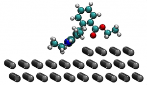 Both the Marcus Theory model of electron transfer and the Landauer approach to conductance in molecular junctions are successfull for describing interfacial behavior at the semi-quantitative level. Similarly, old but useful model treatments such as Poole-Frenkel or space charge limited currents arguments are often helpful for qualitatively understanding the dynamics of these processes. But in connection with electrocatalysis, photoelectron catalysis, ultrafast measurements, and nanoscale structure-function relationships, these approximations may fail, even qualitatively! The MURI team works on developing an entirely new picture for nanoscale (and atomic scale) charge injection, based on the time-dependent Schrodinger equation, using modeled geometries and Green’s function techniques. This advance is now reasonable, because of the very powerful new computational techniques.
To our knowledge, nothing like this approach has ever been tried on the molecule/electrode interface dynamics previously, but such calculations have proven very valuable in other applications such as photoexcited states of molecules. The time-dependent approach permits understanding single-molecule electrochemical processes for any energy, in a time-dependent fashion – this helps controlling effects like overvoltage and coherent multichannel tunneling, and actually seeing (from the simulations) the dynamical effects of interfacial geometry on transport.
Area 2 – Nanoscale scanning electrochemical microscopy (SECM)
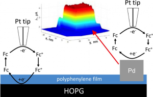
Reference: Blanchard et al. Langmuir 2016, 32 (10), pp 2500–2508
There have been several reports involving nanoscale electrodes and imaging with the scanning electrochemical microscope (SECM). For example, Bard and Mirkin showed trapping of single molecules with a nanoelectrode in a nanoscale interelectrode space. Imaging of DNA and proteins with this resolution was demonstrated by controlling the humidity above a mica electrode so that the tip could be controlled with this thin layer. A number of reports using cantilevers with electroactive tips have also appeared. However, none of these have demonstrated the simplicity of tip preparation and instrument control needed to make the technique widely applicable to broader scientific studies, as we propose in the following sections. We envision that nanoscale SECM and the nanoelectrodes that we develop will find extensive use in ultrafast electrochemistry, single nanoparticle electrochemical studies, mapping of surface catalytic heterogeneity, biological imaging, intracellular electrochemical experiments, and many other areas.
Topic areas include:
- Development of Nanoelectrode Probes (1–10 nm) for Nanoscale SECM
- NanoSECM Intrument Development
- Characterization of Tips and Verification of Instrument Properties
- Approach curves, Fits to Theory – Diffusion
- Approach curves, Fits to Theory – Kinetics
- Imaging Known Defined Structures on the Nanoscale
Area 3 – Nanopore Systems for Delivery of Single Molecules and Nanoparticles to Electrochemical Interfaces
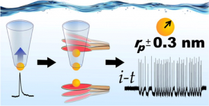 Reference: German et al. ACS Nano, 2015, 9 (7), pp 7186–7194
The MURI team works on integrating solid-state nanopores with nanoscale SECM (Area 2), creating new methodologies for simultaneous delivery and investigation of single redox molecules and electrocatalytic particles. The overarching scientific goal is interfacing single-molecule electrochemistry and spectroscopy with emerging biophysical single-molecule nanopore methods to create feedback control systems for controlling and studying electrochemical behavior. This unique new capability will allow: 1) controlled delivery of finite numbers of molecules/nanoparticles to a solid/liquid interface; 2) investigation of the transport and electrontransfer dynamics of single molecules and particles; and 3) solution-phase patterning of surfaces with single molecules and electrocatalytic nanoparticles. Nanopore and protein ion channel stochastic analyses have undergone significant advances in the past decade, largely driven by applications as single-molecule sensors, for structural analysis and sequencing of DNA, and by the use of solid-state nanopores as nanoparticle Coulter counters. The MURI team employs nanopores in an entirely new application: the controlled delivery of single molecules and particles to a solid/liquid interface.
Area 4- Multimodal Electrochemical Imaging
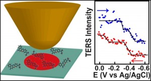
Reference: Kurouski & Mattei et al. Nano Lett., 2015, 15 (12), pp 7956–7962
In this research goal, SECM is interfaced with optical imaging techniques, which provides complementary information on the heterogeneity of various electrochemical systems at the molecular level. We are developing instrumentation in which SECM is integrated with single molecule tip-enhanced Raman scattering (SMTERS) and super-resolution fluorescence imaging. Application of these multi-modal approaches will be used to study 1) single molecule spectroelectrochemistry; 2) electrocatalysis of single metal nanoparticles; and 3) biomolecules at interfaces.
