ILD Round-up 8-4-23

This week in ILD conference, Tim presented a case of fibrotic interstitial lung disease in a middle-aged woman who was born and raised in India and has lived in the US for several years. The CT showed diffuse fibrotic changes with air trapping but no particular apicobasilar gradient. CHP was suspected but HP panels negative and no clear exposure had been identified.

Dr. Parekh inquired whether the patient ever participated in preparation of chapati, which Tim & Anthony had not yet discussed with the patient. On follow up with the patient, she has made chapati every day of her adult life. Interestingly, she had not been involved in chapati baking in last 4-6 months because of hand arthritis. Interestingly, this time course correlated with a previously unexplained improvement in her symptoms.


I. What is the typical radiographic pattern associated with hypersensitivity pneumonitis?

Key point: hallmark of HP is air trapping – expiratory imaging as obtained in HRCT is essential for detection. Air trapping (hypoattenuation), alongside ground glass and normally perfused unaffected lung produces the pathognomonic “three density” or “head cheese” CT finding in HP
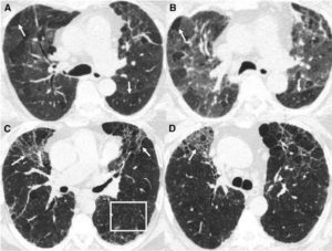
Figure: https://www.atsjournals.org/doi/10.1164/rccm.201608-1675PP

Non-fibrotic Hypersensitivity Pneumonitis: groundglass opacities in random axial and craniocaudal distribution; small (<5mm) centrilobular nodules, air trapping/mosaic attenuation.

Fibrotic Hypersensitivity Pneumonitis: similar distribution and pattern with interposed reticulation, traction bronchiectasis +/- honeycombing. Fibrosis tends to spare basilar lung zones, distinguishing from UIP pattern

HP diagnostic approach:

II. Besides exposure remediation, what is the standard pharmacologic treatment for CHP?

Key point: immunosuppression is the mainstay of pharmacologic treatment for hypersensitivity pneumonitis with evidence of active inflammation

  • Initial tx with corticosteroid
    • 0.5-1 mg/kg/d pred or equivalent, taper to 20 mg/d within 3 mo with close follow-up of PFTs, 6MWD and clinical response
  • When to consider cytotoxic agent (MMF, AZA)
    • Up front with steroids if antigen not readily identifiable
    • Maintenance if response to steroids depending on remediation of antigen, side effect tolerance
  • Antifibrotic treatment in patients with chronic fibrosing ILDs with a progressive phenotype (FDA approved based on INBUILD data)


III. What is “baker’s lung” and in whom should we suspect it? How does this differ from hypersensitivity pneumonitis from occupational exposure to flour?

Baker’s Lung: One of the most reported occupational lung diseases in western countries. It is characterized by respiratory symptoms such as airflow obstruction and bronchial hyper-responsiveness that has been shown to develop in work environments in which there is continued exposure to bakery flour dust.

  • Suspect Baker’s lung in anyone with an occupational or recreational exposure to large quantities of freshly baked breads (bakers, pastry chefs, confectioners, etc.) and asthma-like symptoms.
  • Baker’s lung is caused by Aspergillus-derived carbohydrate cleaving enzymes (ex: alpha-amylase) commonly used to enhance baked products.

Hypersensitivity pneumonitis: As a separate entity, Hypersensitivity pneumonitis has been reported in occupational exposure to bread flour, related either to precipitins against A fumigatus itself (a common culprit for HP in farmers lung), the flour mite Acarus siro, or weevil infestation.


Works Cited:

https://doi.org/10.1378/chest.13-1734 https://www.atsjournals.org/doi/full/10.1513/AnnalsATS.202009-1195CME https://www.atsjournals.org/doi/10.1164/rccm.201608-1675PP https://www.ncbi.nlm.nih.gov/pmc/articles/PMC4278578/#R1 https://www.atsjournals.org/doi/full/10.1164/rccm.202005-2032ST https://www.sciencedirect.com/science/article/abs/pii/009167499290470M


ILD Roundup 11/11/22

It’s been a few weeks since our most recent ILD roundup – we’re glad to be back! This weeks ILD conference was chock full of pearls as usual.

1. First, we discussed the implications of leukocyte telomere length (LTL) testing on decision to use immunomodulatory therapy. Recall that PANTHER-IPF showed evidence of harm in patients with IPF receiving prednisone, azathioprine and n-acetylcysteine (NAC).


Could LTL serve as a biomarker to predict patients at risk of harm from use of immunomodulatory therapy in IPF?   

This question was asked in a recent post-hoc analysis1 of the PANTHER-IPF2 and unpublished ACE-IPF study. The authors found that short LTL (<10%ile) was associated with an increased risk of the composite outcome of death, lung transplantation or FVC decline in those exposed to prednisone/azathioprine/NAC (HR 2.84; 1.02-7.87, p=0.04). This association was not found in either cohort when patients with LTL >10%ile were examined.  

The authors propose that this may be related to unmasking of an immune dysfunction phenotype in patients with short LTL through immunosuppression. When the same criteria were applied to an unrelated cohort of patients participating in a longitudinal observational study at UTSW, there was actually a significant improvement in the prednisone/azathioprine/mycophenolate group with LTL >10%ile.   

Kaplan-Meier curve – UTSW cohort


2. Our next patient was a woman in her 70s with GERD and chronic joint pain. with CT imaging after mechanical fall concerning for ILD. Has developed progressive DOE over past year, with steroid responsiveness. The overall CT pattern was most consistent with fibrotic NSIP, but perilobular opacities were also noted. A differential consideration of organizing pneumonia3 was discussed. 


What is a perilobular opacity?  

A perilobular opacity refers to polygonal opacity around interlobular septa and with sparing of the secondary pulmonary lobule. As Dr. Agrawal brought up to the group, this tends to have more diffuse distribution than a a “reversed halo/atoll sign” which is a focal finding.   

“Reverse halo”, or “Atoll” sign in organizing pneumonia


Perilobular opacity in association with bronchial wall thickening and bronchial dilation in organizing pneumonia


What are the radiographic features most consistent with organizing pneumonia, and what are their primary differential considerations?  


3. A final case we discussed was a former tobacco user in his 70s, with RA on MTX, Humira and prednisone, former asbestos exposure, who presented to VA clinic with progressive DOE over past 6-12 mo. A transbronchial biopsy performed in 2021 with negative cytology for malignancy but otherwise non-diagnostic. CT with showed significant asbestos related pleural disease. Reticulation was seen mostly in association with pleural plaques. Despite the diagnosis of seropositive RA, our multi-disciplinary consensus was asbestos-related pulmonary fibrosis. The question of anti-fibrotic treatment was raised.  


What is the evidence for antifibrotic therapy in asbestos-related pulmonary fibrosis?  

Remember, the INBUILD4 trial showed evidence of benefit (lower annual rate of FVC decline) for antifibrotics in non-IPF fibrosing ILDs. Did they include asbestos-related fibrosis? Hard to say! Looking at the supplementary information (see Table below), 81/663 patients fell into category of “other ILDs” which did include exposure-related ILDs among others, but didn’t specifically mention asbestosis.  


The RELIEF5 study was a phase II placebo controlled RCT that looked at use of antifibrotic agents for non-IPF ILDs (fibrosing NSIP, CHP, and asbestos related pulmonary fibrosis). Patients enrolled had experienced disease progression despite conventional therapy. Of note, only 5 of 127 patients included with asbestos-related pulmonary fibrosis. They followed patients for 48 weeks and reported a significantly lower rate of decline in FVC as a % of predicted value.   

The annual rate of decline in FVC (-36.6 vs –114.4, p=0.21) did not meet statistical significance. Why is this relevant? A quick refresher6 on the endpoints for the IPF anti-fibrotic trials:  


Sources:


ILD Roundup 10/19/22

At ILD conference this week, a patient with progressive RA-ILD was discussed. A change in the patient’s rheumatoid arthritis medication was to be determined with her rheumatologist, but she was also recommended to start nintendanib

I. What is the evidence for anti-fibrotic therapy outside of IPF?

Prior to 2019, the efficacy of antifibrotic therapy in non-IPF fibrosing lung disease was unknown. INBUILD was a double-blind, placebo controlled RCT to investigate the efficacy of antifibrotic therapy in non-IPF fibrosing lung disease.

Let’s approach this study using the PICO framework!

Population:

All patients had to meet criteria for progression of ILD in the past 24 months despite treatment with an FVC =< 45% and DLCO <80%.

Breakdown of population by diagnosis:

·


Intervention:

Nintedanib 150mg BID

Comparison:

Placebo

Outcomes:

  • Primary endpoint=annual rate of decline in FVC
  • The annual rate of decline in FVC was significantly lower in patients who received nintedanib than those who received placebo.
  • Diarrhea was a common adverse event

The patient population was stratified by ILD with or without a UIP pattern of fibrosing ILD. Nintendanib decreased the rate of decline in FVC regardless of the pattern of fibrosis in this patient population.

In another case, our thoracic radiologist Dr. Parekh pointed out an example of dendritic pulmonary ossification.

II. What is dendritic pulmonary ossification (DPO)?

  • Chronic, progressive metaplastic ossification of lung parenchyma
  • Pattern of ossification resembles the dendrite of a neuron

Top: neuron with dendrites
Bottom: coronal view of CT showing dendritic pulmonary ossification

  • Dendritic pulmonary ossification is a rare condition (1.6/1000 autopsies) and is associated with IPF, ARDS, COPD, organizing pneumonia, rare earth pneumoconiosis, asbestosis, heavy metal exposure, and chronic aspiration.
  • Many cases are idiopathic and several weak associations with other non-pulmonary diseases have been reported in the literature.


Sources:

  1. Flaherty et al. N Engl J Med 2019;381:1718-27. DOI: 10.1056/NEJMoa1908681
  2. Fernández-Bussy et al. Respiratory Care April 2015, 60 (4) e64-e67; DOI: https://doi.org/10.4187/respcare.03531
  3. Reddy TL, von der Thüsen J, Walsh SF. . Idiopathic Dendriform Pulmonary Ossification. Journal of Thoracic Imaging. 2012; 27 (5): W108-W110. doi: 10.1097/RTI.0b013e3182326c38.