Elderly immunocompromised man with several years of intermittent dry cough but more green and productive over the last month. Carries diagnosis of asthma on symbicort and PE on eliquis. Afebrile, satting well on room air, but some bibasilar crackles.
Chest imaging showed scattered opacities, slightly reticular, more at the bases. He improved with CTX/azithro; after a week, grew a variety of NTM organisms from multiple samples, including M. abscessus (usually want to treat, more likely to progress and more virulent than MAC).
Group consensus was to send for susceptibilities at National Jewish, but not to treat given symptomatic improvement and repeat CT was much improved (near normal) without treatment for NTM.
Teaching point: Susceptibility showed azithromycin susceptible but high MIC of 32, concerning for something that would have inducible azithro resistance. Functional erm gene also a marker of difficulty to eradicate.
Ongoing discussions regarding treating (if so, with what? to what goal?) and immunosuppression adjustments…
Thank you Dr. Olson for a fantastic morning report!
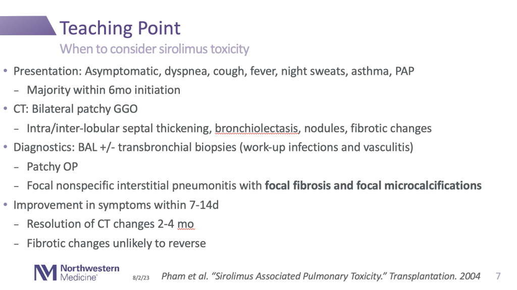
A middle-aged man s/p transplant on sirolimus and tacrolimus presents with progressive dyspnea and fevers.
Chest CT with progressive ground glass opacities in bilateral upper lobes that has progressed now to extensive cystic/cavitary disease over the last few months, despite antibiotics.
BAL with positive histo antigen in serum, urine, BAL, pleural fluid, and on Karius testing! Improves with holding immunosuppression and itraconazole treatment.
This case may have been infectious, but thank you for the great teaching point on when to consider sirolimus toxicity:
Today in Pulmonary Report, the Emily Olson series continues with a case of cystic lung disease. Today, Emily described the management of a young woman with recurrent secondary spontaneous pneumothorax, found to have cystic lung disease.
Before we dive into today’s report, we encourage you to take this opportunity for some retrieval practice from prior NU PCCM blog posts:
For review, here are the blog posts associated with polls above
A case of cystic lung disease in patient with Sjogren’s syndrome from ILD Roundup last July.
Management of secondary spontaneous pneumothorax with delayed resolution (Leak for a week!) from Emily’s Morning Report last August
A case of secondary spontaneous pneumothorax from cystic lung disease from Ale’s Morning Report last September
Alright! Now that the knowledge is fresh, let’s dive in! First, we discussed definitional criteria and a differential diagnosis (1) of cystic lung disease:
Another outstanding quick reference on this topic is Nick Mark’s OnePager (2):
Emily outlined the pragmatic 5-step approach described by Raoof et al.
Further refinement of differential involves considering the apicobasilar gradient of illness:
Emily’s case presented a bit of a diagnostic dilemma – LAM was clinically suspected but VEGF testing performed and unrevealing. Patient ultimately proceeded to VATS resection of a peripheral cyst/bulla, but pathology was inconclusive. This brought up an important discussion point – does it really matter if we know that cause?
Revealing cause of cystic lung disease may have important management implications for our patients:
Genetic inheritance of cystic lung diseases and multi-system illnesses associated. Genetic counseling/sequencing may be indicated
Differential considerations for cystic lung diseases include metastatic malignancies and certain infectious processes with specific management
Dr. Russell also brought up the higher risk of malignancy in patients with Birt-Hogg-Dube (BHD) specifically – particularly colorectal cancer. Recall also from Ale’s morning report – 7x risk of RCC, screening with annual ultrasound in US. BHD, which follows an AD inheritance pattern, may be confirmed by genetic testing revealing a mutation in the FLCN gene, although testing may not be covered by insurance.
Dr. Singer also mentioned the NEJM study (3) that found sirolimus to be associated with stabilization of lung function and improved quality of life in patients with Lymphangioleiomyomatosis (LAM).
Today in report, Emily @EmilyOlsonMD discussed a great case an individual with incidentally discovered pulmonary sequestration who had presented with progressive weakness, and for whom there was concern for malignancy & paraneoplastic syndrome.
First, we discussed a radiographic differential diagnosis:
Emily next took us ALL the way back to medical school embyrology to discuss the two mechanisms of sequestration (intralobar and extralobar)
How does the natural history of intralobar vs extralobar sequestration vary?
Emily’s patient ended up undergoing IR embolization before eventually having a resection of their pulmonary sequestration. Fortunately, the explanted lung tissue did not show malignancy.
Patient in her 60s, former smoker 40py, with abnormal LDCT imaging. Some dyspnea but thought to be related to significant weight gain during pandemic.
Special shoutout to Dr. Agrawal’s Youtube channel and MIPs!
The case had a small endobronchial lesion on CT. We reviewed the differential for endobronchial lesions:
And focused on trahceobronchial tumors, which are rare, 0.6% of pulmonary tumors. (With a fun jeopardy matching series of slides, not captured in this post.)
DIPNECH – associated with bronchial carcinoid, consider in asthma patient with endobronchial abnormality.
Thanks for a great review and a fun interactive session, Dr. Rowe!
On Monday, second year fellow Tom Bolig presented the course of a middle aged undomiciled man with heroin use disorder and recurrent severe asthma exacerbations. This patient had no history of peripheral eosinophilia or IgE elevation. He was non-adherent to maintenance inhaler therapy. He was admitted to the MICU after intubation for asthma exacerbation following unintentional heroin overdose.
This prompted a discussion of the entity of potentially fatal asthma (PFA), defined by Northwestern’s own Paul Greenberger (1,2)
Potentially fatal asthma (PFA) is a clinical condition wherein 1+ of the following are present:
History of endotracheal intubation
Acute respiratory acidosis or respiratory failure from asthma
2+ episodes of pneumothorax or pneumomediastinum from asthma
2+ episodes of acute severe asthma despite long-term use of oral steroids (pre-biologic era) or other asthma medications
Why is this so important?
Condition with high risk for mortality and a young (mean 40 ya) patient population!
Identification may be the first step to tailored management
Loss to follow-up more commonly observed in patients who died of disease
Comorbid psychiatric illnesses and social barriers to health commonly observed
Back to Tom’s patient – a NBBAL was performed with PMN predominance, non-pathologic growth on cx, strongly positive amylase and a galactomannan Ag of 3.87. CT imaging showed patchy bibasilar infiltrates, not consistent with invasive pulmonary aspergillosis (IPA).
What are the most recent recommendations on interpretation of testing in suspected IPA?
All of the following from 2019 ATS Guidelines (3) with strong recommendation/high quality evidence
If hematologic malignancy/solid organ transplant with suspected IPA, obtain serum galactomannan
If serum galactomannan negative in above but high suspicion remains, obtain BAL galactomannan
If serum galactommannan positive but risk factors for false positive (active chemotherapy, suspected/confirmed mucositis), obtain BAL galactomannan
If severe immune compromise as above and suspected IPA, add serum aspergillus PCR to testing above
Tom’s patient fell outside the best studied population (hematologic malignancy and transplant) for galactomannan testing for IPA, and suspicion for disease based off of CT evidence was low. Although this has not been described in the literature, Ben Singer raised the possibility of aspiration of fungal cell wall contents from oropharynx as a putative cause of transiently elevated BAL galactomannan.
Finally, Tom discussed “Mab” therapy for asthma, providing a quick reference chart that takes some of the guesswork out of determining indications:
Thanks, Tom!
Sources:
Allergy and Asthma Proceedings (1988); 9(2):147-152.
Elderly man with history of coronary disease and heart failure presenting with respiratory failure and moderate large bilateral effusions. On lifelong DAPT.
1) Will thoracentesis help?
Dyspnea from altered respiratory mechanics (pushing down on diaphragm and against chest wall) and impaired gas exchange, hypoxemia through pulmonary shunt v/q mismatch, and thoracentesis often doesn’t have immediate effect on gas exchange.
Small study – shows improves respiratory mechanics and oxygenation.
2) What should we do about AC?
Old guidelines from 2010 recommend against, practice patterns are all over the place. Papers with small numbers all had pretty low bleeding outcomes, especially in the hands of an experienced provider with ultrasound use.
More recently, Patel et al reported 451/8951 thoracentesis done on NOAC/antiplatelets, and there were ZERO bleeding complications. Done by IP.
Interestingly, draining one side led to less fluid on the other wise. Suspected to be Buffalo chest – plero-pleural communication – refers to life-threatening condition in which a simultaneous bilateral pneumothorax occurs due to this communication. Can be seen post-op thoracic surgery too.
Middle aged man with Crohn’s disease referred for cough, mucus production.
CT scan showed diffuse bronchiectasis and narrowed BI.
Bronchiectasis is associated with Crohn’s disease, see Review of pulmonary involvement of Crohn’s – often with suppurative mucus. Unclear if treatment of Crohn’s improves pulmonary features.
Focusing on tracheobronchial stenosis – malignancy, chronic infections, inflammatory (GPA, relapsing polychondritis, associated with IBD, amyloid)
Underwent dilation with IP -> path showing chronic inflammation, cultures negative for infectious organisms -> significant symptomatic improvement.
Thanks, Dr. Ludwig! Happy Halloween!
Amy Ludwig, MD, Pulmonary & Critical Care Medicine
Dr. Lawrence presented a case today of a patient with a 50+ pack-year smoking history with concern for bowel obstruction and need for surgery – imaging showed very significant bulla.
What might you recommend before the surgical procedure?
Optimization of medication (referred to COPD clinic)
Smoking cessation
PFTs (points were brought up about not using DLCO/VA, though might be helpful to compare to TLC; and N2 washout suboptimal in patient with obstruction)
Bullous emphysema – giant bulla is >1cm in diameter and giant bulla occupies at least 30% of hemothorax


 Thanks for an outstanding discussion, Emily!


