ILD Round-up 8-4-23

This week in ILD conference, Tim presented a case of fibrotic interstitial lung disease in a middle-aged woman who was born and raised in India and has lived in the US for several years. The CT showed diffuse fibrotic changes with air trapping but no particular apicobasilar gradient. CHP was suspected but HP panels negative and no clear exposure had been identified.

Dr. Parekh inquired whether the patient ever participated in preparation of chapati, which Tim & Anthony had not yet discussed with the patient. On follow up with the patient, she has made chapati every day of her adult life. Interestingly, she had not been involved in chapati baking in last 4-6 months because of hand arthritis. Interestingly, this time course correlated with a previously unexplained improvement in her symptoms.


I. What is the typical radiographic pattern associated with hypersensitivity pneumonitis?

Key point: hallmark of HP is air trapping – expiratory imaging as obtained in HRCT is essential for detection. Air trapping (hypoattenuation), alongside ground glass and normally perfused unaffected lung produces the pathognomonic “three density” or “head cheese” CT finding in HP
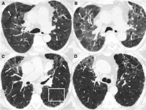
Figure: https://www.atsjournals.org/doi/10.1164/rccm.201608-1675PP

Non-fibrotic Hypersensitivity Pneumonitis: groundglass opacities in random axial and craniocaudal distribution; small (<5mm) centrilobular nodules, air trapping/mosaic attenuation.

Fibrotic Hypersensitivity Pneumonitis: similar distribution and pattern with interposed reticulation, traction bronchiectasis +/- honeycombing. Fibrosis tends to spare basilar lung zones, distinguishing from UIP pattern

HP diagnostic approach:

II. Besides exposure remediation, what is the standard pharmacologic treatment for CHP?

Key point: immunosuppression is the mainstay of pharmacologic treatment for hypersensitivity pneumonitis with evidence of active inflammation

  • Initial tx with corticosteroid
    • 0.5-1 mg/kg/d pred or equivalent, taper to 20 mg/d within 3 mo with close follow-up of PFTs, 6MWD and clinical response
  • When to consider cytotoxic agent (MMF, AZA)
    • Up front with steroids if antigen not readily identifiable
    • Maintenance if response to steroids depending on remediation of antigen, side effect tolerance
  • Antifibrotic treatment in patients with chronic fibrosing ILDs with a progressive phenotype (FDA approved based on INBUILD data)


III. What is “baker’s lung” and in whom should we suspect it? How does this differ from hypersensitivity pneumonitis from occupational exposure to flour?

Baker’s Lung: One of the most reported occupational lung diseases in western countries. It is characterized by respiratory symptoms such as airflow obstruction and bronchial hyper-responsiveness that has been shown to develop in work environments in which there is continued exposure to bakery flour dust.

  • Suspect Baker’s lung in anyone with an occupational or recreational exposure to large quantities of freshly baked breads (bakers, pastry chefs, confectioners, etc.) and asthma-like symptoms.
  • Baker’s lung is caused by Aspergillus-derived carbohydrate cleaving enzymes (ex: alpha-amylase) commonly used to enhance baked products.

Hypersensitivity pneumonitis: As a separate entity, Hypersensitivity pneumonitis has been reported in occupational exposure to bread flour, related either to precipitins against A fumigatus itself (a common culprit for HP in farmers lung), the flour mite Acarus siro, or weevil infestation.


Works Cited:

https://doi.org/10.1378/chest.13-1734 https://www.atsjournals.org/doi/full/10.1513/AnnalsATS.202009-1195CME https://www.atsjournals.org/doi/10.1164/rccm.201608-1675PP https://www.ncbi.nlm.nih.gov/pmc/articles/PMC4278578/#R1 https://www.atsjournals.org/doi/full/10.1164/rccm.202005-2032ST https://www.sciencedirect.com/science/article/abs/pii/009167499290470M


ILD Round-up 3-3-23

Last week, Anthony presented a patient with a reduction in DLCO and mild restrictive spirometric defect on PFTs CT scan showed questionable interstitial changes. We discussed further evaluation and management of ILAs. 

How do we define Interstitial Lung Abnormalities (ILAs) (1)? 

  1. incidentally identified  
  1. non-dependent interstitial changes (reticulation, groundglass, traction bronchiectasis, honeycombing, non-emphysematous cysts) involving  
  1. at least 5% of a lung zone  
  1. in patients for whom ILD is not clinically suspected 

Mimickers of ILAs include dependent atelectasis, interstitial edema, aspiration (GGOs, TiB opacities) and others 

Image: focal paraspinal fibrosis (arrow) in contact with spinal osteophytes, a mimicker of ILAs


How common are ILAs, and what is their significance? 

  • Large population-based (MESA, AGES, FHS, Nagano) and smoking/lung ca. screening (ECLIPSE, NLST, COPDGene, MILD) cohorts found that ILAs are present in 4-10% of individuals screened. 
  • Mean age of scan with ILA varies between 62-78 
  • Progression to ILD observed in 20% at 2 years, and 45-65% at 4-6 years (depending on study, not all cohorts assessed for progression) 
  • Mortality risk associations suggested in several studies, with RR between 1.3-2.7 (95% CI >1, depending on study, not all cohorts assessed for mortality) 
  • Risk factors for ILAs are similar to ILD: increased age, male sex, tobacco smoke exposure 

Table: clinical risk factors for ILD progression (2)


Can ILAs be further categorized, and what are implications of these categories? 

Non-subpleural: usually non-progressive and not associated with increase in mortality 


Subpleural non-fibrotic: sub pleural distribution associated with higher rates of mortality


Subpleural fibrotic: lower zone predominant, reticulation, traction bronchiectasis associated with 6x increased risk of progression   


How should we manage patients with ILAs? 

An algorithmic approach is described in the image below:  


Takeaway points:

  • ILAs are (1) incidentally found (2) non-dependent (3) involving at least 5% of lung zone and (4) in patients for whom ILD is not suspected
  • ILAs are present in 4-10% of patients screened, and typically found in 6th/7th decade of life, and are associated with increased mortality (RR 1.3-2.7)
  • Progression is 20% at 2 years, 45-65% at 5 years 
  • Risk of progression is higher in subpleural location and if reticulations/fibrosis are present

Sources: 

  1. PMID: 32649920 / DOI: 10.1016/S2213-2600(20)30168-5
  2.  PMID: 34374589 /  DOI: 10.1148/radiol.2021204367 


ILD Round Up 2-19-23

In last week’s ILD conference, we discussed a patient with seropositive RA for whom RA-ILD was suspected.  

First, a quick reminder of ILD patterns associated with rheumatoid arthritis (1) from July ILD Round Up: 

Dr. Rishi Agrawal mentioned that this patient’s HRCT pattern was most consistent with UIP; however, given this patient’s history of connective tissue disease, “exuberant honeycombing” may be expected, and the lack of any honeycombing was somewhat atypical.  

What characteristics are commonly associated with a UIP pattern in patients with CT-ILD, and how might they differ from UIP in a patient with IPF? 

A 2018 study (2) looked at patients with UIP with either a diagnosis of IPF or CTD-ILD. Exuberant honeycombing was found in only 6% of IPF patients compared to 22% of patients with CTD-ILD.  

While we’re on the subject…what is exuberant honeycombing? 

Exuberant honeycombing (3) refers to extensive honeycomb-like cyst formation within the lungs comprising >70% of the fibrotic portions of the lungs.

Finally, we discussed the patient’s prior treatment regimen, which included methotrexate (MTX).  

What is the likelihood of methotrexate-associated interstitial lung disease in patients with RA-ILD on methotrexate? 

A case-control study (4) looking at the association between MTX exposure and ILD (n=1083 patients)  found an inverse correlation between MTX exposure and RA-ILD (OR 0.46: 0.24-0.90, p=0.02). Another meta-analysis (5) of 30 studies with 15000 total patients found no association (RR 1.02: 0.73-1.44) for non-infectious adverse respiratory events with MTX use.  

Dr. Carrie Richardson noted that many patients with more severe RA will have already failed therapy with methotrexate for their articular symptoms, which may confound anecdotal reports of ILD associated with MTX use.  

Dr. Jane Dematte also mentioned the distinct entity of MTX associated hypersensitivity. This association is well-recognized, with frequency level 5 from Pneumotox profile corresponding to >200 reported cases in the literature.  

The actual frequency of occurrence depends on definitional criteria (see figure below) but is likely <1%. Below, see the commonly used Carson Criteria for diagnosis of MTX associated pneumonitis (MTX-P). 

Sources: 

1. DOI: 10.1183/09059180.00008014 / PMID: 25726549 

2. DOI: 10.2214/AJR.17.18384 / PMID: 29140119 

3. DOI: 10.1016/j.ejro.2022.100419 / PMID: 35445144 

4. DOI: 10.1183/13993003.00337-2020 / PMID: 32646919 

5. DOI: 10.1093/rheumatology/kez337 / PMID: 31504978 


ILD Roundup 11/11/22

It’s been a few weeks since our most recent ILD roundup – we’re glad to be back! This weeks ILD conference was chock full of pearls as usual.

1. First, we discussed the implications of leukocyte telomere length (LTL) testing on decision to use immunomodulatory therapy. Recall that PANTHER-IPF showed evidence of harm in patients with IPF receiving prednisone, azathioprine and n-acetylcysteine (NAC).


Could LTL serve as a biomarker to predict patients at risk of harm from use of immunomodulatory therapy in IPF?   

This question was asked in a recent post-hoc analysis1 of the PANTHER-IPF2 and unpublished ACE-IPF study. The authors found that short LTL (<10%ile) was associated with an increased risk of the composite outcome of death, lung transplantation or FVC decline in those exposed to prednisone/azathioprine/NAC (HR 2.84; 1.02-7.87, p=0.04). This association was not found in either cohort when patients with LTL >10%ile were examined.  

The authors propose that this may be related to unmasking of an immune dysfunction phenotype in patients with short LTL through immunosuppression. When the same criteria were applied to an unrelated cohort of patients participating in a longitudinal observational study at UTSW, there was actually a significant improvement in the prednisone/azathioprine/mycophenolate group with LTL >10%ile.   

Kaplan-Meier curve – UTSW cohort


2. Our next patient was a woman in her 70s with GERD and chronic joint pain. with CT imaging after mechanical fall concerning for ILD. Has developed progressive DOE over past year, with steroid responsiveness. The overall CT pattern was most consistent with fibrotic NSIP, but perilobular opacities were also noted. A differential consideration of organizing pneumonia3 was discussed. 


What is a perilobular opacity?  

A perilobular opacity refers to polygonal opacity around interlobular septa and with sparing of the secondary pulmonary lobule. As Dr. Agrawal brought up to the group, this tends to have more diffuse distribution than a a “reversed halo/atoll sign” which is a focal finding.   

“Reverse halo”, or “Atoll” sign in organizing pneumonia


Perilobular opacity in association with bronchial wall thickening and bronchial dilation in organizing pneumonia


What are the radiographic features most consistent with organizing pneumonia, and what are their primary differential considerations?  


3. A final case we discussed was a former tobacco user in his 70s, with RA on MTX, Humira and prednisone, former asbestos exposure, who presented to VA clinic with progressive DOE over past 6-12 mo. A transbronchial biopsy performed in 2021 with negative cytology for malignancy but otherwise non-diagnostic. CT with showed significant asbestos related pleural disease. Reticulation was seen mostly in association with pleural plaques. Despite the diagnosis of seropositive RA, our multi-disciplinary consensus was asbestos-related pulmonary fibrosis. The question of anti-fibrotic treatment was raised.  


What is the evidence for antifibrotic therapy in asbestos-related pulmonary fibrosis?  

Remember, the INBUILD4 trial showed evidence of benefit (lower annual rate of FVC decline) for antifibrotics in non-IPF fibrosing ILDs. Did they include asbestos-related fibrosis? Hard to say! Looking at the supplementary information (see Table below), 81/663 patients fell into category of “other ILDs” which did include exposure-related ILDs among others, but didn’t specifically mention asbestosis.  


The RELIEF5 study was a phase II placebo controlled RCT that looked at use of antifibrotic agents for non-IPF ILDs (fibrosing NSIP, CHP, and asbestos related pulmonary fibrosis). Patients enrolled had experienced disease progression despite conventional therapy. Of note, only 5 of 127 patients included with asbestos-related pulmonary fibrosis. They followed patients for 48 weeks and reported a significantly lower rate of decline in FVC as a % of predicted value.   

The annual rate of decline in FVC (-36.6 vs –114.4, p=0.21) did not meet statistical significance. Why is this relevant? A quick refresher6 on the endpoints for the IPF anti-fibrotic trials:  


Sources:


ILD Roundup 10/19/22

At ILD conference this week, a patient with progressive RA-ILD was discussed. A change in the patient’s rheumatoid arthritis medication was to be determined with her rheumatologist, but she was also recommended to start nintendanib

I. What is the evidence for anti-fibrotic therapy outside of IPF?

Prior to 2019, the efficacy of antifibrotic therapy in non-IPF fibrosing lung disease was unknown. INBUILD was a double-blind, placebo controlled RCT to investigate the efficacy of antifibrotic therapy in non-IPF fibrosing lung disease.

Let’s approach this study using the PICO framework!

Population:

All patients had to meet criteria for progression of ILD in the past 24 months despite treatment with an FVC =< 45% and DLCO <80%.

Breakdown of population by diagnosis:

·


Intervention:

Nintedanib 150mg BID

Comparison:

Placebo

Outcomes:

  • Primary endpoint=annual rate of decline in FVC
  • The annual rate of decline in FVC was significantly lower in patients who received nintedanib than those who received placebo.
  • Diarrhea was a common adverse event

The patient population was stratified by ILD with or without a UIP pattern of fibrosing ILD. Nintendanib decreased the rate of decline in FVC regardless of the pattern of fibrosis in this patient population.

In another case, our thoracic radiologist Dr. Parekh pointed out an example of dendritic pulmonary ossification.

II. What is dendritic pulmonary ossification (DPO)?

  • Chronic, progressive metaplastic ossification of lung parenchyma
  • Pattern of ossification resembles the dendrite of a neuron

Top: neuron with dendrites
Bottom: coronal view of CT showing dendritic pulmonary ossification

  • Dendritic pulmonary ossification is a rare condition (1.6/1000 autopsies) and is associated with IPF, ARDS, COPD, organizing pneumonia, rare earth pneumoconiosis, asbestosis, heavy metal exposure, and chronic aspiration.
  • Many cases are idiopathic and several weak associations with other non-pulmonary diseases have been reported in the literature.


Sources:

  1. Flaherty et al. N Engl J Med 2019;381:1718-27. DOI: 10.1056/NEJMoa1908681
  2. Fernández-Bussy et al. Respiratory Care April 2015, 60 (4) e64-e67; DOI: https://doi.org/10.4187/respcare.03531
  3. Reddy TL, von der Thüsen J, Walsh SF. . Idiopathic Dendriform Pulmonary Ossification. Journal of Thoracic Imaging. 2012; 27 (5): W108-W110. doi: 10.1097/RTI.0b013e3182326c38.


ILD Roundup 10/11/22

This week, we discussed the case of a 48 yo with chronic cough who presented to care for evaluation of abnormal CT chest, which showed multifocal peripheral and peribronchovascular pulmonary nodules.

I. How can we use distribution of nodules to narrow differential diagnosis? 

Back to our patient! Using our knowledge and the peribronchovascular/subpleural distribution of nodules, a focused differential diagnosis was discussed (sarcoidosis, silicosis/coal-workers pneumoconiosis, lymphangitic carcinomatosis). Absent occupational exposures were noted, and lack of associated adenopathy/effusions/extrathoracic disease was discussed. Bronchoscopy with EBUS/TBNA and TBBx was performed. A representative sample of the transbronchial biopsy is shown below:

Transbronchial biopsy showing tightly packed non-caseating granulomas with partial hyalinization in a peribronchial distribution compatible with nummular sarcoidosis


Based on the CT and biopsy findings, the group arrived upon a diagnosis of nummular sarcoidosis. Given relatively mild symptoms and normal pulmonary function tests, Dr. Russell suggested the use of high dose inhaled steroids.  

II. What is the role of inhaled corticosteroids (ICS) in the management of sarcoidosis?

The rationale for use? Sarcoidosis is a disease process that follows a lymphatic distribution in the lungs – ICS targets the endobronchial lymphatics. In short, the data is limited! A Cochrane review of corticosteroid use in sarcoidosis included 7 studies that assessed ICS (budesonide 800-1600 mcg/d or fluticasone 880-2000 mcg/d) use, specifically in patients with stage 1 & 2 disease. No improvements in CXR or PFTs were observed, although one study showed a modest improvement in DLCO/VA and others reported improvement in symptom scores.  

III. What are the extra-pulmonary screening recommendations in sarcoidosis? 

Sources:

  • Chest 2006; 129(3):805-15. (link) 
  • Cochrane Database Syst Rev. 2005; 2005(2): CD001114. (link) 
  • AJRCCM 2020; 201(8):e26-e51. (link) 

ILD Roundup – 10/4/22

It’s been awhile since our last ILD round-up, but we are glad to be back in action! This week we talked about a young man with CVID, ITP and persistent groundglass opacities. We discussed a concern for granulomatous and lymphocytic interstitial lung disease (GL-ILD)


I. What is GL-ILD?  

Seen in patients with CVID. With the advent of effective therapies (namely IVIG), increased prevalence of non-infectious complications of CVID (non-infectious complications now seen in 70% of patients with CVID).  

GL-ILD is seen in 8-20% of cases of CVID, making it the most common ILD in this condition. It may also be seen in CTLA-4 deficiency. Associated with splenomegaly, immune-mediated cytopenias, and ITP. BAL demonstrates increased percentage of CD21lo B cells. Pathology demonstrates a peribronchiolar lymphocytic infiltrate, usually associated with granulomas (94% in one case series) and often with organization.  

Image: peribronchial and interstitial lymphocytic infiltration

Image: Epithelioid granuloma


II. What are the characteristic HRCT findings of GL-ILD? 

Solid & subsolid nodules, groundglass opacities, reticulations, mediastinal and hilar adenopathy. Less likely to contain reticulation, TBE, honeycombing, masses or consolidation. A radiologic DDX includes infection, organizing pneumonia, LIP, sarcoidosis and lymphoma. As alreadt noted, splenomegaly (bottom image) is common.

III. How is GL-ILD treated? 

Consensus guidelines with strong agreement that mainstay of therapy is optimization of IVIG therapy. Whether to proactively treat or enter active surveillance after IVIG optimization is less well established, nor is whether antibiotic prophylaxis (as is often the case in CVID) is warranted. Corticosteroids are frequently used, but without clear evidence basis or consensus. Potential second line agents include azathioprine, rituximab, MMF.  

Sources:

  1. J Allergy Clin Immunol Pract. (2017); 5:938-945. (link) 
  2. Front Immunol. (2021); 12:627423. (link) 
  3. Hum Pathol (2016); 46(9): 1306-1314. (link) 
  4. https://radiopaedia.org/cases/granulomatous-lymphocytic-interstitial-lung-disease?lang=us  

ILD Round Up – 9/2/22

This week, a 75-year-old woman former smoker with history of recurrent PEs with abnormal CT imaging was presented. Her work up was notable for a +ANA (1:640) and an HP panel with low-level positive mold antibodies. A TTE showed normal LV and RV size and function with a mildly elevated RVSP. Her high-resolution chest CT had evidence of prominent mosaic attenuation, peripheral and peribronchovascular reticulations, ground glass, traction bronchiectasis, and extensive air trapping. Her PFTs normalized with a course of prednisone except for a persistent, mildly reduced DLCO. The patient had improvement in her cough and SOB but still had spells of lightheadedness. The question presented to the group was “Is there a need for further diagnostics for HP or pulmonary hypertension?”


I. What is mosaic attenuation?

Mosaic attenuation on CT is a heterogeneous pattern of attenuation that resembles…a mosaic.

This differing attenuation may represent:

(a) patchy interstitial disease

(b) diffuse ground glass disease (think acute pulmonary edema, viral/atypical pna, DAH)

(c) obliterative small airways disease

(d) occlusive vascular disease (aka “mosaic oligemia”)

(e) combination of any of the above


Differential for mosaic attenuation secondary to small airways disease:

In this patient, the presence of mosaic attenuation could represent either HP, mosaic oligemia from pulmonary vascular disease, or both.


II. Is there a way to differentiate between etiologies of mosaic attenuation radiologically?

  • As mentioned by Dr Rishi Agrawal, one of the best ways to distinguish small airways disease from other forms of mosaic attenuation is by looking at your expiratory imaging on HRCT.
  • In non-airways-related causes of mosaic attenuation, the lungs should increase in attenuation on expiratory imaging diffusely. This contrasts with what you should see in small-airways disease, where gas trapping will accentuate differences in attenuation.


III. Takeaways

  • Mosaic attenuation is a non-specific finding on CT that can represent disease of the small airways, interstitium, alveoli, or pulmonary vasculature.
  • In an undifferentiated patient, it is important to consider mosaic oligemia 2/2 PAH as a cause of mosaic attenuation.
  • Pulmonary diseases affecting the small airways includes a broad differential (table above for reference).
  • If the attenuation diffusely increases on expiration, it suggests that the etiology of the mosaic attenuation is NOT related to the small airways.


References:

Fleischner Society: Glossary of Terms for Thoracic Imaging https://doi-org.turing.library.northwestern.edu/10.1148/radiol.2462070712

Mosaic Attenuation: Etiology, Methods of Differentiation, and Pitfalls https://doi.org/10.1148/rg.2015140308

ILD Roundup 8/19/22

I. A patient with prior clinical diagnosis of pulmonary sarcoidosis presented to care at NMH with progressive pulmonary fibrosis. A transbronchial biopsy with EBUS was performed. The radiographic pattern and biopsy results were thought to be inconsistent with sarcoidosis; a differential of indeterminate UIP v NSIP (idiopathic v IPAF) remained. We discussed sending the patient for Envisia testing.


What do we mean when we say “Envisia”?

  • Envisia Genomic Classifier
    • Developed using machine learning applied to bulk RNA sequencing data from lung biopsy in combination with histologically-confirmed diagnoses
    • Helps differentiate UIP from non-UIP histologic patterns by transcriptomic signature

Envisia was validated in the BRAVE (Bronchial Sample Collection for a Novel Genomic Test) studies


How well can Envisia distinguish UIP from other pathology in conventional TBBx?

  • Envisia identified UIP in transbronchial biopsy with a specificity of 88% and sensitivity of 70%
  • Among patients who had “possible or inconsistent UIP” on HRCT, Envisia showed 81% positive predictive value for biopsy-proven UIP


Liu et al. BMJ 2022;377:e066354 https://doi.org/10.1136/bmj-2021-066354

Raghu et al. Lancet Respir Med 2019; 7: 487–96 10.1016/S2213-2600(19)30059-1


II. A patient who is undergoing evaluation for transplant had chronic hypersensitivity pneumonitis (cHP) in his differential diagnosis. We discussed the predictive value of BAL lymphocyte count given the fibrotic/chronic nature of his disease


What is the predictive value of BAL lymphocytosis in cHP?

  • A 2020 ERJ systematic review and meta-analysis suggested an association between BAL lymphocytosis and fibrotic cHP
    • BAL lymphocyte percentage higher in cHP (42.8%, CI 37.7-47.8) compared to IPF (10%, CI 6.9-13.1) and other IIPs
    • Analysis demonstrated that a BAL lymph% cutoff >20% optimized sensitivity and specificity for cHP compared to other IIPs

  • 2018 ATS/ERS/JRS/ALAT IPF diagnostic guidelines conditionally recommend BAL in patients with suspected IPF and a non-diagnostic HRCT pattern
    • A meta-analysis of eight studies in the most recent guidelines found no difference in BAL lymphocyte percentage between IPF and cHP

ILD roundup 8/12/22

I. A 62 yo female with family history of pulmonary fibrosis with an NSIP pattern

What is the relationship between telomere length and familial pulmonary fibrosis?

-Heterozygous mutations in coding regions of two telomerase genes (TERT :: protein component, TERC :: RNA component) have been associated (found in 15% of kindreds) with familial IPF  

Pulmonary fibrosis is seen in 50% of women and 60% of men 60 years and older with TERT mutations


-UIP radiograph pattern (74% HRCT) and UIP pathologic pattern (86% surgical lung biopsy) is most commonly encountered 

-Even in sporadic cases of IPF, a significantly higher proportion of individuals have telomere lengths <10th percentile compared with controls, even absent TERT/TERC mutations (present in 1-3% of sporadic cases) 


Plotting mean telomere length against age with 10-90th percentile in normal controls mapped in blue region


https://www.atsjournals.org/doi/pdf/10.1164/rccm.200804-550OC 

https://doi.org/10.1371/journal.pone.0010680 

II. 69 yo F with seropositive RA and progressive fibrosing ILD, found to have upper zone predominant fibrosis, honeycombing, TBE.  

What HRCT & histopathologic patterns are most commonly associated with RA-ILD?
 -UIP pattern on HRCT most commonly encountered, between 40-60% of cases.  

-Histopathology usually correlates with HRCT. MC encountered histopathologic patterns are UIP, NSIP, OP 


Aggregated data from multiple studies demonstrating the most common histopathologic patterns in CTD-ILDs. Note larger proportion of UIP in RA-ILD


-RA-ILD associated with UIP pattern has an increased risk of disease progression and death, and increased risk of flares compared with RA-ILD with NSIP pattern 

Kaplan-Meier curve showing RA-ILD with UIP pattern; cumulative survival intermediate between RA-ILD with NSIP and IPF (another ILD characterized by UIP HRCT and histopath)


What is the role of antifibrotic therapy in non-IPF ILD? 

– A recent trial (INBUILD – 2019) looked at nintedanib (antifibrotic FDA approved for IPF) in patients with non-IPF fibrosing interstitial lung disease (n=663) over 12 months of follow-up 


Figure demonstrating the primary endpoint, decline in FVC over period of followup (188 mL in placebo versus 80 mL in nintedanib group).


-As a secondary endpoint, mortality was less in intervention group (11.5% placebo vs  8.1% nintedanib) although this was not statistically significant 

– Since publication of INBUILD trial, FDA has approved use of nintedanib/Ofev for non-IPF progressive fibrosing ILDs 

Finally, for a current and comprehensive review of the treatment of fibrosing interstitial lung disease including IPF and other progressive pulmonary fibrosis (PPF), make sure to check out a recent review in BMJ by our very own Gabby Liu, Scott Budinger and Jane Dematte!  

An algorithmic approach to diagnosis and management


https://doi.org/10.1378/chest.09-0444 

https://www.nejm.org/doi/full/10.1056/NEJMoa1908681 

https://www.bmj.com/content/377/bmj-2021-066354 

https://www.fda.gov/news-events/press-announcements/fda-approves-first-treatment-group-progressive-interstitial-lung-diseases