“Outcomes associated with corticosteroid dosage in critically ill patients with acute exacerbations of chronic obstructive pulmonary disease,” AJRCCM, 2016

“Outcomes associated with corticosteroid dosage in critically ill patients with acute exacerbations of chronic obstructive pulmonary disease,” AJRCCM, 2016, University of Colorado

Question: Is high-dose or low-dose systemic corticosteroid therapy better for intensive care unit (ICU) patients with acute exacerbations of COPD (AE-COPD)?

 Study Type: Retrospective cohort study using a large national quality and health care use database.

Study Population: Patients >40 years of age admitted to an ICU and treated with systemic corticosteroids by hospital day 2 for AE-COPD.  Patients excluded if steroid dose >1g/day methylpred, OSH tx, re-admission within 30 days of a prior discharge, solid organ transplant, or presence of pulmonary embolism, pneumothorax, shock, or antifungal therapy.

Study Groups: Patients divided into high-dose corticosteroids (>240 mg/day methylpred) and lower-dose corticosteroids (≤240 mg/day methylpred, i.e., 60 mg Q6 hrs or less).

Primary Outcome: In-hospital mortality

Results: 17,239 patients included in analysis.  After propensity score matching, there was no difference in the primary outcome between groups.  Patients in the lower-dose group had better clinical outcomes (less time on mechanical ventilation, less time in the ICU, less time in the hospital) and less steroid-related complications (decreased insulin requirements and fewer fungal infections).

Caveats: Retrospective design, association ≠ causation, and potential for unmeasured confounding.

Take-home Point: High-dose systemic corticosteroid therapy does not improve outcomes for critically ill patients with AE-COPD and is associated with a higher rate of steroid-related complications.

The third international consensus definitions for sepsis and septic shock (sepsis-3)

“The third international consensus definitions for sepsis and septic shock (sepsis-3),” JAMA 315: 801, 2016, SCCM sepsis definitions task force

Intro:

  • initial definitions of sepsis, severe sepsis, and septic shock from 1991; defined severe sepsis as sepsis complicated by organ dysfx which could progress to septic shock, defined as hypotension despite adequate fluid resuscitation; revisited in 2001
  • the definitions of sepsis, severe sepsis, and septic shock have remained largely unchanged for more than 2 decades

Process of developing new definitions:

  • recognizing the need to reexamine current definitions, the euro society of critical care medicine and the society of critical care medicine convened a task force (TF) of 19 experts in critical care, ID, surgery, and pulmonology (no EM) in jan 2014
  • existing definitions were revisited in light of an enhanced appreciation of the pathobiology and the availability of large EMR databases and pt cohorts
  • expert consensus process forged agreement on updated definitions and the criteria to be tested in the clinical arena
  • the agreement between potential clinical criteria and the ability of the criteria to predict outcomes (eg ICU admission, mortality) were then tested in multiple large EMR databases
  • systematic lit review/metaanalysis and delphi consensus methods were also used for the definition and clinical criteria describing septic shock
  • when compiled, the TF recommendations were circulated to major international societies and other relevant bodies for peer review and endorsing (a total of 31 endorsing societies) (again, no EM)

 Challenges and opportunities:

  • because no gold standard diagnostic tests exist for sepsis, the TF sought definitions and supporting clinical criteria that were clear and fulfilled multiple domains of usefulness and validity
  • the original conceptualization of sepsis as infection + ³2 SIRS criteria focused solely on inflammatory excess; sepsis is now recognized to involve early activation of both pro- and anti-inflammatory responses, along with major modifications in nonimmunologic pathways such as cardiovascular, neuronal, autonomic, hormonal, bioenergetic, metabolic, and coagulation, all of which have prognostic significance; a broader perspective also emphasizes the significant biological and clinical heterogeneity in affected individuals
  • the current ³2 SIRS criteria to identify sepsis was unanimously considered by the TF to be unhelpful
  • the “sequential organ failure assessment” (SOFA) score looks at severity of organ dysfx via the following variables: P/F ratio (resp), platelets (coag), bili (liver), BP (cardiovascular), GCS (neuro), creat and UO (renal); a higher SOFA score is associated with increased mortality, but outside the critical care community the score is not well known + cumbersome to use
  • multiple definitions for septic shock are currently in use resulting in significant heterogeneity in reported mortality
  • the public needs an understandable definition of sepsis and health care providers require improved clinical prompts and diagnostic approaches to facilitate earlier identification and an accurate quantification of the burden of sepsis

Results/recommendations:

Definition of sepsis:

  • sepsis is defined as life-threatening organ dysfx caused by a dysregulated host response to infection
  • under this definition (“life-threatening organ dysfx”), the term “severe sepsis” becomes superfluous

Clinical criteria to identify pts with sepsis:

  • the TF evaluated which clinical criteria best identified pts most likely to have sepsis; this objective was achieved by interrogating large datasets of hospitalized pts with presumed infection, assessing agreement among existing scores of inflammation (SIRS) or organ dysfx (SOFA), and delineating their correlation with subsequent outcomes; in addition, multivariable regression was used to explore the performance of 21 bedside and lab criteria proposed by the 2001 TF
  • the EMR included 150,000 pts with suspected infection (from 12 hosps within the univ of pittsburgh system); two outcomes – ICU stay ³3 days and mortality – were used to assess predictive validity
  • for ICU pts, a change in SOFA score ³2 from baseline was superior to SIRS criteria; the TF recommends the use of SOFA score ³2 from baseline to identify life-threatening organ dysfx and, thus, sepsis
  • for non-ICU pts, the TF found that 2 or more of the following clinical variables – altered mental status, syst BP £100, and RR ³22 – offered good predictive validity; this new measure is termed qSOFA (for quick SOFA) and provides simple bedside criteria to identify life-threatening organ dysfx and, thus, sepsis; the TF recommends that qSOFA criteria be used to prompt clinicians to further investigate organ dysfx, to initiate or escalate therapy as appropriate, and to consider referral to critical care specialists
  • thus, for ER pts, the TF recommends the following for identification of pts with sepsis: ³2 of the qSOFA criteria: altered MS, syst BP £100, RR ³22

Definition of septic shock:

  • septic shock is a subset of sepsis in which underlying circulatory and cellular metabolic abnormalities are profound enough to substantially increase mortality

Clinical criteria to identify septic shock:

  • 3 variables were identified to test in cohort studies – MAP <65, lactate >2, and need for vasopressor(s) to maintain MAP ³65 after volume resuscitation
  • the first database interrogated was the surviving sepsis campaign’s international multicenter registry; a total of 20,000 septic pts; the mortality for septic pts with all 3 variables was >40%
  • these same 3 variables were then used to interrogate 2 unrelated large EMR datasets – univ of pittsburgh (12 hosps, 6,000 pts) and kaiser (20 hosps, 54,000 pts); the mortality for septic pts with all 3 variables was 35-55%
  • thus, the TF recommends the following criteria for identification of pts with septic shock: MAP <65, lactate >2, and need for vasopressor(s) to maintain MAP ³65 after volume resuscitation

Controversies – lactate levels:

  • because lactate offered no meaningful change in the predictive validity beyond ³2 of the qSOFA criteria in the identification of pts with sepsis, the TF could not justify adding the complexity/cost of lactates alongside these simple bedside criteria
  • the TF recommendations should not, however, constrain the monitoring of lactate as an indicator of illness severity or as a guide to therapeutic response
  • some TF members suggested that elevated lactate represents an important marker of “covert shock” in the absence of hypotension

Implications:

  • simple clinical criteria (qSOFA) that identify pts with sepsis (ie pts with evidence of infection who are likely to have a prolonged ICU course +/- death) have been developed and validated
  • there is potential conflict with current organ dysfx scoring systems, early warning scores, ongoing research studies, and pathway developments

“Editorial: new definitions for sepsis and septic shock: continuing evolution but with much still to be done,” wake forest

  • the TF assessed the predictive validity of SOFA, SIRS, and qSOFA in a primary cohort that included 150,000 pts with suspected sepsis and a confirmatory analysis that included 700,000 pt encounters at 156 US and non-US hosps
  • for identifying pts with sepsis: the investigators found that in the ICU the best predictive value was found with change in SOFA score >2 from baseline; in non-ICU settings, the best predictive value was found with the qSOFA score
  • for identifying pts with septic shock: the TF conducted a systematic review and metaanalysis of 92 studies informing a delphi process that created the new definition, then tested the variables identified by the delphi process in cohort studies using datasets from the surviving sepsis registry, univ of pittsburgh hosps, and kaiser hosps
  • according to the new recommendations:
    • sepsis is now identified by evidence of infection + life-threatening organ dysfx, clinically characterized by an acute change in the SOFA score ³2 from baseline (ICU pts) or ³2 of the 3 clinical variables of the qSOFA score (nonICU pts)
    • septic shock is now identified by MAP <65, lactate >2, and the need for vasopressor(s) to maintain MAP >65 after volume resuscitation
  • there is no longer any mention of the SIRS criteria (HR >90, RR >20, T >38° or <36°, WBC >12K or bands >10%)
  • there remain concerns with the quality of the information used to generate the updated criteria
  • regarding the new qSOFA score: because this score was retrospectively derived from databases that had substantial gaps in clinical info for pts treated outside ICUs, qSOFA will require prospective real-world validation before it should enter routine clinical practice

Prevalence of pulmonary embolism among patients hospitalized for syncope

“Prevalence of pulmonary embolism among patients hospitalized for syncope,” NEJM, 2016, Italy

Question: What is the prevalence of pulmonary embolism in patients hospitalized for a first episode of syncope?

Study Type: Multicenter cross-sectional study at 11 hospitals in Italy

Study Population: Patients admitted for syncope were eligible. Exclusion criteria included pregnancy, previous episodes of syncope, and use of anticoagulation.

Study Groups: Enrolled patients had a simplified dichotomized Wells score calculated and a D-dimer drawn. Patients with a Wells score ≤4 and a negative D-dimer were considered to have PE excluded. Patients with a Wells score >4, a positive D-dimer, or both underwent further evaluation for PE with either a PE-CT or V/Q scan

Primary Outcome: Presence of a pulmonary embolism on either PE-CT or V/Q scan.

 Results: Of 2,584 patients who visited the ED for syncope, 717 were admitted, and 560 (22% of all patients) were included in the study.  The median age of the cohort was 80. 330 patients (60%) had a PE excluded by a low-risk Wells score and a negative D-dimer.  PE was identified in 97 of the remaining 230 patients (17% of the study cohort).  Of patients diagnosed with a PE, 45% had a RR>20, 33% had a HR >100, 40% had clinical signs of DVT, and 20% had active cancer.  17 patients with a PE (18%) had a small thrombus burden identified by either a subsegmental PE on CT-PE or a perfusion defect of 1-25% on V/Q scan.

Caveats: No information on outcomes of patients discharged from the ED, normal diagnostic testing for PE risk stratification (troponin, BNP, echo) not included, age-adjusted D-dimer not used, no outcomes information for patients diagnosed with a PE, concern regarding generalizability (the study cohort was elderly and the prevalence of PE in Europe is different than in the United States).

Take-home Point: A diagnosis of pulmonary embolism should be considered in patients admitted to the hospital with a first episode of syncope.

Commentary: There is concern that this article will be used to justify indiscriminate testing for PE in patients with syncope (one educational blog review of this article is entitled “The Impending Pulmonary Embolism Apocalypse”).  My thoughts on what this trial should and should not do:

  1. This trial should remind clinicians of the important Christopher study (JAMA 2006) which showed that a clinical decision score (a dichotomized Wells score) combined with D-dimer testing can exclude PE and avoid unnecessary CT scans with a low miss rate (<0.5%) in a large number of patients. Interns and residents not familiar with this article should give it a read. 
  1. This trial should also remind clinicians that pulmonary embolism is a potential cause of syncope and that especially in older patients with concerning signs/symptoms, a diagnostic evaluation for VTE should be considered. Of patients diagnosed with PE in this trial, 45% were tachypneic, 33% were tachycardic, 40% had signs of DVT, and 40% had a high risk Wells score on presentation all in a cohort in which 20% had active cancer. Considering the diagnosis of VTE in this group seems like a reasonable thing to do.  It is notable that 25% of patients who were diagnosed with a PE did not have clinical manifestations of VTE. 
  1. The prevalence of 17% found in this study should not be generalized to practice in theUS. Multiple studies have found the prevalence of PE in Europe to be consistently higher than in the United States (around 25-30% for Europe vs. <10% in the US).  The patients in this trial were also elderly and likely a higher risk cohort than other syncope populations.  The striking difference between the prevalence of 17% found in this trial and the very low prevalence found in the studies they cite (<3%) likely in part reflect their rigorous testing and unique patient cohort.
  2. Identification should not be conflated with causation. 18% of patients had a small thrombus burden identified on either CT or V/Q scanning. It is not clear from the trial if these small clots were the cause of syncope or if identifying and treating them impacts patient outcomes.  ​

Levosimendan for the prevention of acute organ dysfunction in sepsis

“Levosimendan for the prevention of acute organ dysfunction in sepsis,” NEJM, 2016, UK

Background: Levosimendan is a calcium-sensitizing drug marketed under the trade name Simdax that is approved in Europe and South American for use in acute decompensated heart failure. It increases cardiac contractility through calcium sensitization of troponin C and vasodilates by opening potassium channels in vascular smooth muscle cells. It is felt to only minimally increase myocardial oxygen demand. Small trials have suggested that it may improve organ perfusion and hemodynamics in sepsis.

Question: Does the addition of levosimendan to standard care in patients with septic shock reduce the severity of organ dysfunction?

Study Type: Multicenter, randomized, double-blind, placebo-controlled trial in 34 ICUs in the UK.

Study Population: Adult patients who required vasopressor support for ≥4hrs for septic shock were eligible for the trial. Exclusion criteria included >24 hrs of vasopressor support prior to enrollment, ESRD, Child-Pugh C liver disease, history of torsades, DNR, weight >125kg, and pregnancy.

Study Groups: Patients randomized to levosimendan or placebo for 24 hours. Additional inotropic medications were allowed if felt necessary by the treating clinician. Orion Pharmaceuticals provided the study drug but was not otherwise involved in the trial.

Primary Outcome: Mean daily SOFA score while in the ICU for a maximum of 28 days.

Results: 516 patients were randomized. The majority of screened patients were excluded because they were outside the 24hr window since meeting inclusion criteria. Notable baseline characteristics: Caucasian (93%), recent surgery (37%), APACE II (25), lung as primary source of infection (39%), beta-blockers at baseline (19%), mechanical ventilation (81%), dobutamine (9%). There was no difference in the mean SOFA score during ICU stay (6.68 +/-3.96 in the levosimendan group and 6.06 +/- 3.89 in the placebo group). Patients given levosimendan required more norepinephrine, were less likely to be liberated from mechanical ventilation, and more likely to experience SVT. Levosimendan did not improve outcomes in any of the pre-specified subgroups including patients with low cardiac output, impaired O2 delivery to tissue, and those on high-dose vasopressors. There was no difference in any mortality measure. Of note, cardiac index was similar throughout the trial in both groups (largely between 3.1-3.5 L/min/m2).

Caveats: Mostly Caucasian patients, does not answer the question of whether levosimendan may be helpful in the initial resuscitation of septic shock (median time from shock to randomization in this trial was 16 hrs), does not answer the question of whether a supranormal cardiac index is helpful in septic shock (both groups had a similar CI and very few patients in the intervention arm would have fit into the “cardiac index” arm of the 1995 NEJM trial we reviewed where CI goal was >4.5), does not answer the question of whether an inotrope is helpful in septic shock as the addition of levosimendan was offset in part by increased dobutamine use in the control arm, cardiac index was only measured in a subset of patients (30%) and measured using a variety of methods (PiCCO, esophageal Doppler, PAC) raising questions about generalizability and accuracy, more dobutamine use in the standard care arm then is seen at NMH.

Take-home Point: The addition of levosimendan to standard care in patients with septic shock does not improve outcomes and may be harmful.

Commentary: For me, an odd hypothesis to think that levosimendan would provide much physiologic benefit in this patient group: resuscitated septic patients who had been on vasopressors for a median of 16 hours and had adequate DO2 (as assessed by a median ScVO2 of 75 at the time of enrollment) especially as another inotrope was allowed in both arms. I would have been more interested in a protocol focused on early resuscitation, perhaps specifically in patients with a low ScVO2 or known cardiac dysfunction comparing levosimendan to placebo (without additional dobutamine) or a three-armed trial with levosimendan vs dobutamine vs placebo (as levosimendan’s ability to only minimally raise myocardial O2 demand offers some potential advantages over dobutamine). In any case, this trial, and others strongly suggest that a supranormal DO2 (or attempts to achieve one) are of little benefit in patients with sepsis. This trial is also a good reminder to take systematic reviews of small trials with a big grain of salt (see “Levosimendan reduces mortality in patients with severe sepsis and septic shock: a meta-analysis of randomized trials,” J Critical Care, 2015).

Azithromycin for acute exacerbations of asthma: The AZALEA randomized clinical trial

“Azithromycin for acute exacerbations of asthma: The AZALEA randomized clinical trial,” JAMA IM, 2016, United Kingdom

Question: Does azithromycin improve outcomes for patients with acute asthma exacerbations?

Study Type: Multicenter, double-blind, placebo-controlled randomized trial

Study Population: Adults with a documented history of asthma who required IV or oral steroids for an acute asthma exacerbation were eligible.  Notable exclusion criteria included the use of oral or intravenous corticosteroids within the previous 28 days and need for intensive care unit admission.

Study Groups: Patients were randomized to receive azithromycin 500mg daily or placebo for 3 days.

Primary Outcome: Diary card summary symptom score at day 10

ResultsOf the 4,500 patients screened, 45% were excluded because they had already received antibiotics.  199 patients were randomized.  Asthma symptom scores measured at day 10 did not differ between the two groups. Similarly, the addition of azithromycin did not improve quality-of-life scores, lung function measurements, or time to 50% reduction in symptom score.

Caveats: Study did not meet enrollment goal and was underpowered, investigators enrolled a select group of patients that treating clinicians felt would not benefit from antibiotic therapy (the 55% of patients not excluded for already receiving abx) perhaps skewing the trial toward a negative result.

 Take-home Point: Azithromycin does not improve outcomes for patients with acute asthma exacerbations.  The trial also shows that abx are still widely prescribed for asthma exacerbations despite not being supported by guidelines or evidence.

Impact of thrombolytic therapy on the long-term outcome of intermediate-risk pulmonary embolism

“Impact of thrombolytic therapy on the long-term outcome of intermediate-risk pulmonary embolism” JACC, 2017, Europe

Question: Does thrombolytic therapy for submassive PE impact long-term outcomes?

Study Type:  Long-term follow-up of a multicenter, double-blind, placebo-controlled trial

Study Population: Study focused on a subset of patients from the PEITHO trial (NEJM, 2012). In PEITHO, 1,006 adults with confirmed PE + RV dysfunction (on either TTE or CT) + elevated troponin I or T were randomized to heparin +/- a single weight-based intravenous bolus of tenecteplase.  After a protocol amendment, consent was obtained from a subset of these patients to obtain 2-year survival data and prospectively conduct long-term clinical and echocardiographic follow-up.

Primary Outcome: Death from any cause at 2 years

Results: 709 patients included (70% of entire PEITHO cohort). Compared to patients without 24-month follow-up, patients in the study had a lower mean body weight (81.8 kg vs. 84.4 kg) and less prior VTE (25% vs. 32%). Patients in the follow-up cohort had a mean age of 66.6 and 8.2% had active cancer. Overall, treatment with tenecteplase was not associated with any improvement in long-term outcomes. Notable results (tenecteplase % listed first): death from any cause between randomization and 2-year follow-up (20.3% vs. 18%), persistent clinical symptoms (36% vs. 30%), NYHA class III or IV dyspnea (12% vs. 10.9%), 1 or more echo indicators of pulmonary HTN or RV dysfunction (44.1% vs 36.6%), PASP (31.6 vs. 30.7)

Caveats: Only included a subset of the PEITHO cohort, only 40% of included patients had TTE f/u, up to 33% of patients had missing data for various endpoints, outcomes not stratified by submassive risk (i.e., rising troponin, progressive hypoxemia, etc), duration to determine risk of CTEPH may not be long enough to truly identify people who develop the disease.

Take-home Point: Among patients with submassive PE (defined by the presence of RV dysfunction and elevated troponin), systemic thrombolytics does not appear to improve long-term mortality, functional outcomes, or rates of pulmonary hypertension compared to anticoagulation alone.

Commentary

–       Despite the limitations noted above (I am not sure why assessing long-term outcomes was not a pre-specified aim of the study before it started), this is an important article.

–       The PEITHO study remains the highest quality trial of thrombolytics for submassive PE. In that trial, lytics decreased the risk of a composite endpoint of hemodynamic collapse or death within 7 days (3% with lytics vs. 6% with placebo driven almost entirely by a decrease in hemodynamic collapse) at the cost of an increased risk of bleeding including ICH (2% with lytics vs 0.2% with placebo for ICH). Important also to recognize that the inclusion criteria for PEITHO were VERY liberal – all you needed was a +trop and some evidence of RV dysfunction. Many would argue that a large # of PEITHO pts should never have been given lytics at all.

–       Based on very flawed studies (TOPCOAT, MOPPET), many clinicians give lytics for submassive PE with the goal of improving functional status and decreasing the risk of CTEPH. The results of this article should temper enthusiasm for such a strategy as there was no signal in any of the outcomes assessed that lytics was beneficial.

–       This study does not specifically address catheter-directed therapy nor does it help answer whether lytics are beneficial in particularly high-risk patients (rising troponins, mild hypoxemia, relative hypotension, rising lactate, etc.).

–       In summary, there is potentially (likely?) a role for lytics in a very high-risk group of patients with submassive PE but accumulating evidence suggests that when lytics are given to all patients who meet criteria for submassive PE, they increase rates of bleeding and provide little short or long-term benefit.

Quantifying unintended exposure to high tidal volumes from breath-stacking dyssynchrony in ARDS: the BREATHE criteria,

“Quantifying unintended exposure to high tidal volumes from breath-stacking dyssynchrony in ARDS: the BREATHE criteria,” Intensive Care Medicine, 2016, USA

Note: I am focusing on only one aspect of this paper, see the comments section for more details

Question: Using novel diagnostic criteria, how often does breath-stacking dyssynchrony (BSD) occur in pts with ARDS?

Study Type: Prospective observational study at 2 US hospitals

Study Population: Adult pts with ARDS placed on mechanical ventilation within the previous 24 hours were eligible provided they were ventilated in the assist-control mode. Exclusion criteria included chronic mechanical ventilation, neuromuscular disease compromising spontaneous ventilation, and impending withdrawal of full supportive care

Study Groups: Airflow and pressure were continuously recorded for 72 hours, coded by an analog-digital converter, and read directly into an analysis program. The flow-time waveform was integrated to calculate cumulative tidal volume change over consecutive machine inspiratory cycles. BSD was identified using the BREATHE criteria (consecutive inspiratory cycles + expiratory times <1 sec + expiratory volume between inspiratory cycles at least 2mL/kg PBW less than first inspiratory cycle volume + •120% set inspiratory time {for pressure-targeted breaths} + BSD TV •2mL/Kg PBW above intended TV) (essentially saying the pt inspires twice before fully exhaling the first breath). Medication administration and ventilator management were directed by the ICU team who were blinded to study analysis.

Primary Outcome: BSD as measured by the BREATHE criteria

Results: 33 pts were enrolled. Notable pt characteristics: mean age (57), shock (91%), pneumonia (83%), duration of MV prior to enrollment (16 hrs), death before discharge or day 28 (27%). Vent characteristics: VC+ (volume-targeted pressure control) (79%), VC (typical volume-cycled ventilation with a set flow rate) (21%), set TV (6 mL/kg PBW), RR (25), PEEP (10), FiO2 (50%), P/F (107). 1,841 hrs were recorded and 2,166,076 breaths were analyzed (80% without neuromuscular blockage). Observed BSD frequency was 27 (7-59) breaths/hr, peak hourly BSD frequency was 170 (55-394) breaths/hr. BSD frequency was sustained for more than 60 breaths/hr during 18% (1-37) of hrs recorded without NMB. TV during BSD was 11.3 (9.7-13.3) mL/kg PBW. Peak airway pressure minimally increased with BSD.

Caveats: Small sample size, 743 hours not recorded or included for various reasons, does not establish any connection between BSD and outcomes including ventilator-induced lung injury.

Take-home Point: Among pts with ARDS ventilated in assist-control mode, breath-stacking dyssynchrony occurs and exposes pts to potentially injurious tidal volume.

Commentary

  • The BREATHE criteria are fine, but for residents easier to just remember that BSD is “inspiratory flow triggered before complete expiration” (i.e., the pt is triggering a 2nd breath before fully exhaling the first).

  • I have purposely ignored one of the main aspects of this study: comparing the BREATHE criteria to other methods of measuring BSD. Unless you are really into reading about ventilator dyssynchrony, this part of the paper is less interesting than the more basic message that BSD occurs (sometimes quite often) in AC modes.

  • This paper is a nice reminder that when you are setting a tidal volume in either VC or VC+, this does not guarantee that this is the volume your pt will receive before exhaling. You may think your pt is receiving “lung-protective ventilation,” but frequent episodes of BSD may cause your pt to receive very large TVs (in the above study, an average Vt of 11 mL/kg PBW). These results highlight the importance of looking at ventilator waveforms in your pts and frequently evaluating synchrony.

  • Whether decreasing BSD improves outcomes remains to be seen. This is one of the proposed mechanisms by which early neuromuscular blockade may improve outcomes in ARDS but more studies are needed to prove this connection.


High-impact PCCM Article Summaries: Aggressive or Moderate Fluid Resuscitation in Acute Pancreatitis (The WATERFALL Trial)


De-Madaria et al. Aggressive or Moderate Fluid Resuscitation in Acute Pancreatitis: The WATERFALL Trial. NEJM 2022

Question: Does aggressive fluid resuscitation compared to moderate fluid resuscitation improve clinical outcomes in patients with acute pancreatitis?


Why ask it: How to administer intravenous fluids (IVF) for patients with acute pancreatitis remains a source of debate. Early IVF may improve pancreatic microcirculatory hypoperfusion and help prevent pancreatic necrosis. However, excessive IVF can contribute to complications including respiratory failure and abdominal compartment syndrome.


Intervention: 249 patients in 8 countries presenting to the emergency department with mild acute pancreatitis randomized to aggressive or moderate fluid resuscitation protocols (see comment for important exclusion criteria and details of intervention).


Results (all written as aggressive IVF group vs moderate IVF group):

  • Development of moderately severe or severe acute pancreatitis (primary outcome)
    • 1% vs 17.3% (adjusted RR 1.3; [95% CI, 0.78 – 2.18], p=0.32)
  • Fluid overload during hospitalization (primary safety outcome)
    • 5% vs 6.3% (adjusted RR 2.85; [95% CI, 1.36 – 5.94])
  • No signal of benefit with aggressive IVF across a range of secondary outcomes (select secondary outcomes listed below)
    • Necrotizing pancreatitis
      • 9% vs 7.1% (adjusted RR 1.95; [95% CI, 0.87 – 4.38])
    • Local complications
      • 5% vs 16.5% (adjusted RR 1.28; [95% CI, 0.74 – 2.22])
    • Any organ failure
      • 4% vs 3.9% (adjusted RR 1.23; [95% CI, 0.47 – 3.23])
    • Respiratory failure
      • 4% vs 2.4% (adjusted RR 2.19; [0.63 – 7.64])
    • ICU admission
      • 6% vs 1.6% (adjusted RR 2.71; [95% CI, 0.64 – 11.51])
    • Death
      • 3% vs 0.8% (adjusted RR 3.05; [95% CI, 0.32 – 28.76]
  • The trial was halted by the DSMB at the first interim analysis due to worse safety outcomes in the aggressive IVF group
  • Similar results in prespecified subgroup analyses of patients with SIRS at baseline and those with baseline hypovolemia

Conclusion: In patients with mild acute pancreatitis, aggressive IVF did not improve clinical outcomes and was associated with more fluid overload compared to moderate IVF.

Comments:

  • Acute pancreatitis pathobiology
    • Intra-acinar activation of trypsin causes autodigestive injury to the vascular endothelium, interstitium, and acinar cells with a resulting inflammatory response
    • Acute pancreatitis and sepsis share similar pathobiology including microcirculatory dysfunction, dysregulated inflammatory and coagulation cascades, and the potential for systemic and end-organ complications
  • Central goals of IVF in acute pancreatitis are the correction of hypovolemia and restoration of perfusion to the pancreatic microcirculation
  • WATERFALL was a multi-center, open-label, parallel-group, controlled superiority trial conducted at 18 centers in 4 countries (India, Italy, Mexico, Spain)
  • Many exclusion criteria
    • Moderately severe or severe disease per the Revised Atlanta Classification
    • NYHA CHF II – IV
    • Uncontrolled HTN
    • Hyper or hyponatremia
    • Hyperkalemia
    • Hypercalcemia
    • Life expectancy < 1 year
    • Chronic pancreatitis
    • Chronic renal failure
    • Decompensated cirrhosis
  • Details of interventions (Lactated Ringers used for all)
    • Aggressive-resuscitation group
      • Enrollment
        • Bolus 20 mL/kg, then infusion 3 mL/kg/hr
      • Hour 3 (“safety checkpoint”)
        • Physical assessment to evaluate for signs of volume overload
        • If present, decrease or stop infusion
      • Hours 12, 24, 48, and 72 (“goal-directed therapy checkpoints”)
        • Hypovolemia
          • Bolus 20 mL/kg, then infusion 3mL/kg/hr
          • Additional boluses of 20 mL/kg if low UOP or SBP
        • Normovolemia
          • Infusion 1.5 mL/kg/hr
          • Stop after 48 hrs if oral feeding tolerated for > 8 hrs
        • Suspicion of fluid overload
          • Decrease or stop infusion
          • Infusion stopped after 48 hrs if oral feeding tolerated for > 8hr
    • Moderate-resuscitation group
      • Enrollment
        • 1.5 mL/kg/hr without bolus in pts without hypovolemia
        • If hypovolemia present, bolus 10 ml/kg over 2 hrs then start infusion
      • Hour 3
        • Physical assessment to evaluate for signs of volume overload
        • If present, decrease or stop infusion
      • Hours 12, 24, 48, and 72
        • Hypovolemia
          • Bolus 10 mL/kg, then infusion 1.5 mL/kg/hr
          • Additional boluses of 10 ml/kg if low UOP or SBP
        • Normovolemia
          • Infusion 1.5 mL/kg/hr
          • Stop after 20 hrs if oral feeding tolerated for > 8 hrs
        • Suspicion of fluid overload
          • Decrease or stop infusion
          • Infusion stopped after 20 hrs if oral feeding tolerated for > 8hr
    • Oral feeding started at 12 hrs in both groups if minimal abd pain per the PAN-PROMISE SCORE
    • Fluid overload identified by at least 2 of the following: symptoms, physical signs, and imaging evidence of hypervolemia
  • Notable patient characteristics
    • Age: ~57
    • Gallstone pancreatitis: 61%
    • CAD: 1%
    • Median BiSPAP score: 1
    • 2 or more SIRS: 26%
  • Results of intervention (all written as aggressive IVF group vs moderate IVF group)
    • Median cumulative IVF
      • 12 hrs: 3.4 L vs 1.5 L
      • 24 hrs: 5.4 L vs 3.3 L
      • 48 hours: 7.8 L vs 5.5 L
      • 72 hours: 8.3 L vs 6.6L

My take

  • The trial asks an important and clinically relevant question. As noted in the 2018 American Gastroenterological Association Guidelines on Initial Management of Acute Pancreatitis, there is a paucity of high-quality evidence to inform how and when to administer IVF for patients with acute pancreatitis
  • The intensity of bedside reassessment in the trial (structured safety and goal-directed therapy checks at hours 3, 12, 24, 48, and 72) exceeds what is provided for many hospitalized patients in a real-world setting. The trial therefore likely underestimates the harm associated with aggressive IVF in less monitored settings.
  • By design, the patients in this trial were not that sick. They had minimal co-morbidities and they could not have any organ failures or local/systemic complications related to their acute pancreatitis at the time of enrollment. The results are therefore not generalizable to the care of critically ill patients with acute pancreatitis. Patients who present with severe disease (who may have more pronounced hypovolemia and be at higher risk of progression to necrotizing pancreatitis) may uniquely benefit from IVF. Conversely, those with chronic pulmonary, cardiac, and renal disease are at higher risk of developing clinically significant complications from aggressive IVF. A tough balance.
  • The trial aimed to enroll 744 patients to detect a 10% difference between groups in the development of moderately severe or severe acute pancreatitis assuming an incidence of 35%. Given the lower-than-expected incidence of moderately severe or severe acute pancreatitis during the trial (20% overall) and the early trial termination at an enrollment of 249 patients, the study is underpowered to detect differences in the primary outcome
  • This trial does not inform a safe lower limit for IVF in acute pancreatitis. Do patients really need an infusion of 1.5 mL/kg/hr at days 2 and 3? My guess is no but this trial doesn’t answer that.
  • My simplified view is that we should approach IVF resuscitation in acute pancreatitis much like we do with sepsis (they share many similarities as noted above). IVF in both settings is probably of most benefit when given early and in patients with more severe disease. For the floor patients we evaluate for MICU transfer (worsening disease or organ dysfunction several days into their hospital stay), ongoing high-volume maintenance fluids are likely of little benefit.
  • As in sepsis care, there is not one perfect marker to guide resuscitation in acute pancreatitis. IVF should be guided by serial reassessment of intravascular volume, perfusion pressure, and tissue oxygenation using all of the imperfect tools at our disposal rather than a one-size-fits-all protocol.


HFNC review

Case

A 55 y/o male with a h/o EtOH abuse is intubated and admitted to the MICU for hypoxemic respiratory failure secondary to aspiration PNA.  Three days later, his vital signs have normalized, he is awake and able to follow commands, minimal secretions are noted with suctioning of his ET tube, and his SpO2 is >95% on 40% FiO2 and 5 PEEP.  He passes a 30 minute trial of spontaneous breathing with a T-piece.

Question

Following extubation, should he be given high-flow nasal oxygen to help prevent the need for reintubation?

Evidence

  1. We previously reviewed the FLORALI trial, NEJM 2015 which looked at the up-front use of high-flow nasal cannula (HFNC) in patients with hypoxemic respiratory failure
  1. 313 patients randomized to HFNC vs NIV
    1. HFNC did not lower need for intubation at 28 days
    2. HFNC did lower ICU and 90-day mortality
  2. Interpretation of trial results complicated by significant cross-over in the use of HFNC and NIV between study arms
  1. There is a growing body of literature on the use of HFNC following extubation
    1. We previously reviewed a JAMA 2015 trial looking at the use of HFNC vs NIV in 830 patients at high-risk of reintubation following cardiac surgery
  1. No difference in treatment failure (defined as re-intubation, switch to other study treatment, or premature study discontinuation) between groups
  2. Roughly 14% required re-intubation in each group (no difference)
  • Similar dyspnea and comfort score between groups
    1. Nasal High-flow versus Venturi Mask Oxygen Therapy after Extubation, AJRCCM 2014, Italy
  1. 105 patients with a P/F < 300 following SBT randomized to oxygen via HFNC (flow 50 L/min) or venturi mask for 48 hrs or until ICU discharge (the low P/F perhaps indicative of a group at higher risk of post extubation hypoxemia and need for reintubation)
    1. P/F ratio at 24 hrs (primary end point) significantly higher in HFNC group (287 vs 247). P/F also higher at 36 and 48 hrs in HFNC group
    2. Patient comfort significantly higher in HFNC group
    3. Significantly fewer reintubations in HFNC group (4% vs 21%) although the study was not powered for this outcome
    1. A new trial was just published on the use of HFNC following extubation in patients felt to be at low-risk of post-extubation hypoxemia – Effect of Post-extubation HFNC vs Conventional Oxygen Therapy on Reintubation in Low-Risk Patients, JAMA 2016, Spain
  1. Methods
    1. Inclusion
      1. Pts on mechanical ventilation >12 hrs who passed an SBT and were defined as low risk of reintubation by having all of the following (few patients in our MICU would meet these criteria)
        1. Age <65
        2. Not initially intubated for CHF
  • Absence of mod-severe COPD
  1. APACHE II <12
  2. BMI <30
  3. No known airway problems and low risk of developing laryngeal edema
  • Adequate cough and requiring suctioning <2xs Q8hrs
  • Not difficult to wean
  1. Mechanical ventilation <7 days
  2. <2 co-morbidities
  1. Exclusions
    1. DNR
    2. Trach
    3. Hypercapnia during SBT
  2. Intervention
    1. Immediately following extubation, pts were randomized to either
      1. HFNC x 24 hrs
        1. set at 10L/min and titrated up at 5L/min intervals until pts experienced discomfort
        2. FiO2 titrated to SpO2 >92%.
      2. Conventional oxygen applied through facemask or nasal cannula titrated to SpO2 >92% x 24hrs
  • Primary outcome
    1. Need for reintubation at 72 hrs
  1. Results
    1. 1739 pts ready to be liberated from the vent  527 randomized (vast majority excluded as they were high risk for reintubation)
    2. Patient characteristics
      1. Age ≈51
      2. Neurologic co-morbidity 7.6% in HFNC group and 12.9% in conventional O2 group (only significant baseline difference between the two groups)
      3. Time on vent ≈ 1-2 days
      4. ≈30% admitted with primary neurologic diagnosis and ≈47% had either scheduled or urgent surgery at admission (not a typical MICU population)
    3. Outcomes
  HFNC (264) Coventional O2 (263) P value
All-cause Reintubation at 72hrs 13 (4.9%) 31 (12.2%) .004
Reintubation for respiratory failure 4 (1.5%) 23 (8.7%) .001
Time to reintubation (hrs) 19 (12-28) 15 (9-31) .99
ICU LOS (median) 6 (2-8) 6 (2-9) .29
Hospital mortality 10 (3.8%) 13 (5%) .94
  1. In multivariable analysis, HFNC was independently and inversely associated with all-cause reintubation.
  2. NNT to prevent 1 reintubation = 14
  1. Conclusion
    1. Among patients felt to be at low risk for reintubation, the use of HFNC following extubation vs conventional O2 reduced the need for reintubation at 72hrs with a NNT of 14


Faculty Feedback

I asked Dr. Ben Singer, Assistant Professor of Medicine in the Division of Pulmonary and Critical Care, whether he felt the JAMA trial was practice changing.  A summary of his comments:

  • Important to remember that supplemental O2 is not an entirely benign therapy.  In both animal models and studies of healthy controls, high levels of FiO2 have been found to promote lung injury.  Supplemental FiO2 after extubation should therefore be used thoughtfully.
  • With this in mind, it is important to note that the majority of the patients in the trial were placed on mechanical ventilation for surgical or neurologic diagnoses (only 16% had a primary respiratory reason for mechanical ventilation).  This population is likely at lower risk of lung injury from prolonged high levels of FiO2 than a patient recovering from PNA or ARDS.  This may have biased the trial toward a positive result.  Interestingly, more patients in the conventional O2 arm of the trial had ARDS (4.2% vs 1.5%) – perhaps contributing to the higher rates of reintubation seen in the control arm.
  • As noted above, the patient population in the trial is not representative of patients typically cared for in the MICU.  As a result, the positive results seen with post-extubation HFNC are not necessarily generalizable to MICU patients.
  • There were 7 cases of laryngeal edema necessitating reintubation in the control arm and 0 cases in the HFNC arm.  This is likely just an artifact of randomization as it is hard to imagine HFNC preventing severe laryngeal edema.  If 5 of the severe laryngeal edema patients had been randomized to the HFNC arm, the results of the trial would not have been significant.
  • The curves from Fig 2  look convincing as they immediately separate during the first 24 hrs (when HFNC is being used) and then become essentially parallel suggesting a real benefit from HFNC

  • Important for housestaff to recognize that this trial excluded hypercapnic patients, a group where the evidence supports the up-front use of NIV following extubation.
  • Overall this was a well performed and methodologically rigorous trial.  The results are provocative but limited by the unique patient population (largely healthy neurocritical care and surgical patients).  The trial highlights the need for a large randomized trial of HFNC following extubation in a more inclusive group of MICU patients


Take-home Points

  • The use of HFNC following extubation may reduce the risk of reintubation when compared to conventional O2.  Further trials are needed to clarify which patients stand to benefit most from this therapy.


HFNC review including FLORALI

Case

A 70 year-old male is brought in by ambulance from his nursing home with 2 days of fever, shortness of breath and purulent sputum production.  He is febrile, tachycardic, and tachypneic with an initial SpO2 of 86% on RA which improves to 92% with 5L NC.  On exam he is alert and oriented but appears ill with crackles at his right lung base.  A portable CXR shows a dense right-sided infiltrate.  ABG does not show any evidence of hypercarbia

Questions

What oxygen delivery device would you use to help manage his hypoxemic respiratory failure?  Is there a role for the use of a high-flow nasal cannula (HFNC)?

Physiology and recent trials

  1. Common O2 delivery devices
    1. In general, maximal flow rates of common O2 delivery device are limited by the ability to effectively heat and humidify gas at high flow rates
    2. Nasal cannula
      1. O2 delivery inefficient as O2 flowing through cannula mixes significantly with entrained room air (unable to obtain delivered FiO2 >40%)
      2. High flow rates poorly tolerated as cool dry air irritates the nares
    3. Face mask (Venturi mask)
      1. Can achieve higher flow rates (6-10L/min). Room air entrained through exhalation ports limits maximal FiO2 to 50%
    4. Non-rebreather mask
      1. Includes special valves which limit entrained air allowing delivery of FiO2 near 95%
      2. Maximal flow rates of 10-15L/min
    5. NIV (BiPAP)
      1. Good evidence to support the use of NIV in acute decompensated heart failure and exacerbations of obstructive lung disease
      2. Cumbersome to set up and interface often uncomfortable for patients
  • Mixed data on its efficacy in acute hypoxemic respiratory failure (AHRF)
  1. HFNC
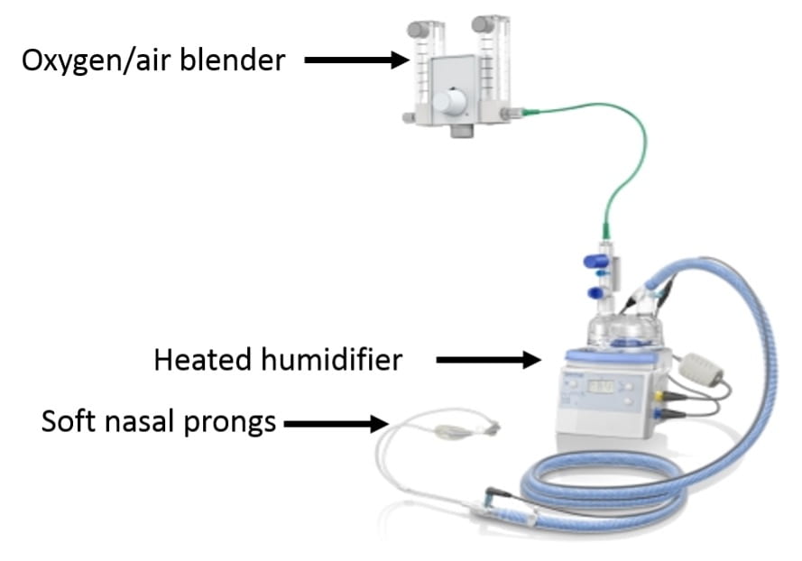
    1. Device (see picture above)
      1. Uses a special oxygen/air blender connected to a heated humidifier to saturate air with water and warm air to body temperature before delivery. This system allows for the delivery of very high flow rates (up to 60L/min)
      2. Connects to the nose with large soft prongs
        1. Potential benefits
          1. Ability to deliver heated and humidified gas at high flow rates
            1. Prevents drying of the airway and interference with mucocilliary clearance
            2. Enhances patient comfort
            3. May help moisten secretions making them easier to clear
            4. May lessen work of breathing by avoiding the bronchoconstricting effects of cold air and lessening the work needed to expectorate secretions
          2. Minimizing entrainment of room air
            1. Patients in respiratory failure often have high flow rates that exceed the flow of nasal cannulas and face-masks resulting in entrained room air which dilutes supplemental O2
            2. Flow rates in HFNC usually exceed patient-generated flows minimizing dilution of delivered oxygen
  • Improved ventilator efficiency
    1. HFNC can continually flush CO2 out of nasopharynx (lowering nasopharyngeal dead space) and allowing more of the minute ventilation to participate in gas exchange
    2. PEEP effect
      1. HFNC increases nasopharyngeal and esophageal pressure approximating levels seen with nasal CPAP
      2. HFNC may therefore provide a small amount of inspiratory assistance, help counter-balance auto-PEEP, and potentially improve oxygenation
    3. Breathing pattern
      1. Some evidence that HFNC helps increase TV and lowers RR
    4. Use
      1. Greatest benefit likely in patients with significant hypoxemia that would ordinarily be given standard high-flow oxygen therapy via a face mask
      2. Avoid in patients with high breathing workloads whose ventilatory failure is worsening (these patients require closely monitored NIV vs intubation)
      3. Usually provides more oxygen and gas flow than is necessary for patients with mild hypoxemia
      4. Initial settings
        1. Initial adjustments should be to flow rate as it is the flow rate that drives the physiologic benefit. Increase flow if RR fails to drop or if breathing remains labored with initial settings
        2. Starting flow rate is usually 35-40L/min
        3. Increasing flow rate should improve FiO2 as the amount of entrained air decreases
      5. There are two recent large randomized trials investigating the use of HFNC which are worth knowing
        1. High-flow oxygen through nasal cannula in acute hypoxemic respiratory failure (FLORALI) – NEJM, 2015
          1. Methods
            1. Multicenter randomized trial in 23 ICUs throughout France and Belgium
            2. Inclusion (need to meet all 4)
              1. RR>25, P/F < 300 while on O2 flow of 10L/min for at least 15 minutes, PaCO2<45, no history of chronic respiratory failure (ie pts with AHRF without concurrent hypercapnia)
            3. Exclusions
              1. PaCO2>45, exacerbation of asthma or chronic respiratory failure, cardiogenic pulmonary edema, neutropenia, shock, GCS <13, contraindications to NIV, urgent need for intubation, DNR (important to exclude obstructive lung disease and decompensated CHF where there is a known benefit to NIV)
            4. Patients randomized to 3 groups
              1. Standard O2 therapy: nonrebreather mask with flow of 10L/min or more with flow adjusted to maintain SpO2 >92% until patient recovered or was intubated
              2. HFNC: flow rate >50L/min and FiO2 100% with FiO2 adjusted to maintain SpO2 >92%. HFNC applied for 48 hours.  In the HFNC and standard oxygen group, a trial of NIV was allowed at the discretion of the MD
              3. NIV: pressure support adjusted to obtain a tidal volume of 7-10cc/kg PBW with initial PEEP between 2-10 cmH20. FiO2 and PEEP then adjusted to maintain SpO2 >92%.  Minimum duration of NIV was 8 hours/day for at least 2 calendar days.  NIV applied during sessions of at least 1 hour and could be resumed if RR was more than 25 and SpO2 <92%.  Between NIV, patients received HFNC
            5. Results
              1. 2,500 patients admitted with HRF  525 eligible  313 randomized (13% of patients with HRF included in trial)
              2. Causes of respiratory failure
                1. CAP: 64%
                2. HAP: 12%
              3. Initial mean settings
                1. Standard O2: O2 flow rate of 14L/min
                2. HFNC: O2 flow rate of 48L/min, mean FiO2 80%
                3. NIV: 8/5, FiO2 70%, TV 9 cc/kg
              4. 25% in NIV group received therapy for <4hrs per day
              5. 40 patients in the HFNC and standard O2 received BiPAP as a rescue therapy
              6. Outcomes (statistically significant results in bold)
HFNC NIV Standard O2 therapy
intubation rate by 28 days (primary outcome) 38% 50% 47%
intubation rate by 28 days in pts with P/F £200 35% 58% 53%
interval between enrollment and intubation 27° 27° 15°
ventilator-free days at day 28 24 19 22
ICU mortality 11% 25% 19%
90-day mortality 13% 31% 22%


  1. 90-day mortality
    1. Hazard ratio for death of 2.01 (1.01-3.99) when comparing standard O2 vs HFNC group and 2.5 (1.31-4.78) when comparing NIV vs HFNC
  2. Intensity of respiratory discomfort and dyspnea score significantly improved in the HFNC group 1 hour after enrollment
  • Conclusions
    1. The use of HFNC vs standard O2 or NIV did not prevent the primary outcome of need for intubation at 28 days
    2. HFNC was associated with several important secondary outcomes
      1. Lower ICU mortality
      2. Lower 90-day mortality
      3. Less need for intubation in patients with a P/F <200
    3. The authors and editorial wonder whether the high tidal volumes achieved with NIV may have contributed to the worse secondary outcomes seen in the NIV group
  1. High-Flow nasal oxygen vs noninvasive positive airway pressure in hypoxemic patients after cardiothoracic surgery – JAMA, 2015
    1. Respiratory failure is common following cardiac surgery with NIV currently the treatment of choice. However, NIV is cumbersome, requires significant resources, and fails in 20% of patients.   Authors wondered whether HFNC may be a better choice.
    2. Methods
      1. Multicenter randomized trial in 6 ICUs throughout France.
      2. Patients (any of the following)
        1. Failure of post-op SBT
        2. Successful SBT in patients with a BMI >30, LVEF <40%, or failure of prior extubations
        3. Successful SBT followed by failed extubation (P/F <300, RR >25 for at least 2hrs, use of accessory muscles)
        4. Exclusions included OSA, tracheostomy, DNI, delirium, nausea, vomiting, altered mental status, hemodynamic instability
      3. Patients randomized to the use of HFNC or NIV
  • Results
    1. 3,217 eligible, 830 randomized (80% were s/p CABG)
    2. Primary outcome of treatment failure (defined as re-intubation, switch to other study treatment, or premature study discontinuation)
      1. 9% in BiPAP vs 21.0% in HFNC (no difference)
      2. No difference in time to treatment failure (1 day in each group)
      3. Roughly 14% required re-intubation in each group (no difference)
      4. No difference in outcome among patients with a P/F < 200 (in contrast to the NEJM study).
      5. Similar dyspnea and comfort score
      6. No difference in ICU or 28 day mortality
    3. Conclusion
      1. HFNC is not inferior to NIV in preventing treatment failure in patients with or at risk for respiratory failure following cardiothoracic surgery


My thoughts

  • I find the results of the FLORALI study less convincing than the associated editorial suggests they are. The significant cross-over between groups (when not on NIV, patients in the NIV group received HFNC and 40 patients not on NIV were placed on NIV as a rescue therapy) and the variable duration of NIV therapy (25% of patients received therapy for <4 hours per day) make a clean comparison between the three O2 delivery methods difficult.  If a patient is on NIV for <4 hours a day and the other 20 hours was managed with HFNC, should their outcome really be associated with NIV use?
  • I also find it difficult to explain the improved 90-day mortality seen with HFNC.  It is hard to picture an interaction between any mode of non-invasive O2 delivery and mortality that does not hinge on precluding the need for mechanical ventilation which was not seen in this trial.  The authors of the trial and the editorial suggest that the high tidal volume achieved with NIV may have worsened lung injury thus leading to more time on the ventilator and increased mortality but it is hard to buy this conclusion if patients managed with NIV did not require intubation any more than patients in the other two groups nor did they have higher rates of refractory hypoxemia.  A trend of P/F ratios in all three arms over time would have been helpful.  Concerns have also been raised about the high rate of septic shock in the NIV arm (31%) vs the HFNC arm (18%)
  • Important to note that 45% of patients required intubation by 28 days. This highlights the importance of closely monitoring patients with AHRF being managed with HFNC or NIV as roughly half will fail. 
  • After reviewing this topic, I find the use of HFNC most appealing in patients who would normally be managed with a higher-flow face mask (venturi or nonrebreather). The ability to deliver very high flows of heated/humidified air seems to offer real physiologic and comfort benefits over typical higher flow devices.
  • We recently discussed the FLORALI study at a pulmonary conference. Faculty raised the important point that for older patients with pneumonia, difficulty with secretion clearance is often what leads to initiation of mechanical ventilation.  In this group of patients, placing them on NIV may inhibit their ability to clear secretions and hasten respiratory failure.  Specifically for this reason, Dr. Wunderink (MICU director) supported the idea of using HFNC over NIV as a first line therapy for older patients with pneumonia and resulting hypoxemic respiratory failure who do not require immediate mechanical ventilation.
  • For those who manage patients with hypoxemic respiratory failure (either in the ER or in the ICU), if you never find yourself initiating therapy with HFNC, you are probably underusing a helpful tool.


Take-home points

  • High flow nasal cannula devices deliver heated and humidified air at very high flow rates. Potential benefits include improved patient comfort, improved secretion clearance, a small PEEP effect, and washout of nasopharyngeal dead space
  • The use of HFNC may improve long-term outcomes in patients with acute hypoxemic respiratory failure although further trials are needed to validate this finding
  • HFNC is not inferior to NIV in preventing treatment failure following cardiothoracic surgery


Attached

  • Helpful review of HFNC from CHEST, 2015
  • FLORALI trial, NEJM, 2015
  • Randomized trial of HFNC following cardiothoracic surgery, JAMA, 2015