Welcome, visitors from Med Ed Day!

We are so excited that Med Ed Day is here!

Northwestern Medical Education (@NU_MedEd) / TwitterJoin us at 8:30 – Short Presentations – Session One – Baldwin Auditorium – as Dr. Rowe presents our preliminary data from the blog and highlights our experiences.

9:00 – Dr. Rowe will present at Eco-Normalization – Searle room

1:45 – Discussing Interactive Asynchronous Learning session – Baldwin Auditorium – a range of experiences with the modern digital age of medical education, including a piece about medical education blogging! Featuring also Dr. Angarone of our friend blog, bit.ly/nuidblog

We’re so glad you’re here, and are always excited to chat about our experience, hear your suggestions, or collaborate!

#NUPCCM Blog Team

Update – thanks to all who came out to support us, and for all of you visiting the blog for the first time after our presentation. We had a great time with the discussion and can’t wait to see the community continue to grow!

From left:
Mike Angarone, Mac Walter, Brianna Valdes, Tim Rowe, Tricia Pendergrast


Morning Report – 9/26/22

Thanks to Dr. Rowe for a great morning report case – a middle aged man with well-controlled HIV, ESRD on HD, referred to clinic for an abnormal CT as part of pre-transplant workup.

CT with some moderate subcarinal and right paratracheal lymphadenopathy, minimal parenchymal findings.

What next?

Differential:

  • Sarcoidosis
  • Infection (TB, endemic fungi, anything really)
  • Malignancy
  • Reactive

EBUS to the rescue! – REMEDY: AJRCCM 2012 study showed that EBUS mediastinoscopies in 87% of cases – 67/77 cases were diagnosed with EBUS; of the 10 undiagnosed, only 6 got diagnoses in mediastinoscopy

Thanks, Tim!

Timothy Rowe, MD, Pulmonary and Critical Care

Academic Half Day 7/21/2022: Acute Hypoxemic Respiratory Failure and ARDS References

Ventilatory Dyssynchrony

Patient-ventilator Interactions: Implications or Clinical Management – AJRCCM 2013

  • Evergreen review of trigger, flow, and cycle dyssynchronies including recognition and management

Alveolar Gas Equation

Teaching an intuitive derivation of the clinical alveolar equations: mass balance as a fundamental physiological principle – Adv Physiol Educ 2020

  • In-depth derivation of alveolar gas equation for those interested in a deep dive

Bias in Pulse Oximetry

Racial bias and reproducibility in pulse oximetry among medical and surgical inpatients in general care in the Veterans Health Administration 2013-19: multicenter, retrospective cohort study – BMJ 2022

  • Recently published large retrospective study that found occult hypoxemia (SaO2 <88% with SpO2 ≥92%) more common in Black vs White patient

Non-invasive Ventilation in the ICU

Philips Respironics V60 User Manual 

  • Critically important to know how to use the devices available to you
  • Page 4-7 reviews trigger, target, and cycle for the different modes available on the V60

ATS/ERS Guidelines on use of NIV for Acute Respiratory Failure – ERJ 2017

  • Consensus guidelines on use of NIV in respiratory failure. A nice summary of the literature on the use of NIV in the pre-intubation and post-extubation setting

Liberation from Mechanical Ventilation in Critically Ill Adults: An Official ATS/ACCP Clinical Practice Guideline – AJRCCM 2017

  • Summary of 3 important guidelines from the ATS/ACCP on evidence-based liberation from mechanical ventilation in critically ill adults. References to the parent documents are provided in this summary

Journal Club: PROSEVA

Prone positioning in severe acute respiratory distress syndrome – NEJM, 2013

  • Landmark RCT which found prone positioning for 16 hrs/day associated with a significant mortality benefit for patients with severe ARDS

Prone position-induced improvement in gas exchange does not predict improved survival in the acute respiratory distress syndrome – AJRCCM 2014

  • Retrospective analysis of PROSEVA data which found that did not find an association between the improvement in gas exchange and survival – suggesting perhaps that proning mechanism of benefit is largely through limiting VILI

Evidence-based ARDS Care

An OfficialAmerican Thoracic Society/European Society of Intensive Care Medicine/Society of Critical Care Medicine Clinical Practice Guideline: Mechanical Ventilation in Adult Patients with Acute Respiratory Distress Syndrome – AJRCCM 2017

  • Evidence-based multi-society guidelines on mechanical ventilation in ARDS. Only LTVV + Pplt < 30 cmH20 and prone positioning for > 12hrs/day in severe ARDS receive strong recommendations FOR

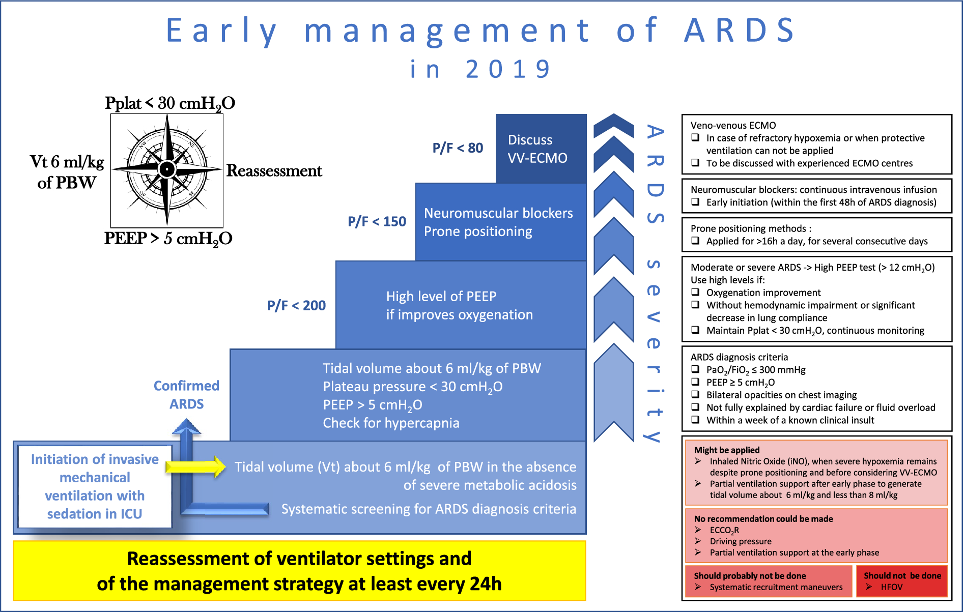
Formal guidelines: management of acute respiratory distress syndrome – Annals Intensive Care 2019

  • More recent guidelines from Société de Réanimation de Langue Française. Their approach:

Fig. 1

High-impact PCCM Article Summaries: Aggressive or Moderate Fluid Resuscitation in Acute Pancreatitis (The WATERFALL Trial)


De-Madaria et al. Aggressive or Moderate Fluid Resuscitation in Acute Pancreatitis: The WATERFALL Trial. NEJM 2022

Question: Does aggressive fluid resuscitation compared to moderate fluid resuscitation improve clinical outcomes in patients with acute pancreatitis?


Why ask it: How to administer intravenous fluids (IVF) for patients with acute pancreatitis remains a source of debate. Early IVF may improve pancreatic microcirculatory hypoperfusion and help prevent pancreatic necrosis. However, excessive IVF can contribute to complications including respiratory failure and abdominal compartment syndrome.


Intervention: 249 patients in 8 countries presenting to the emergency department with mild acute pancreatitis randomized to aggressive or moderate fluid resuscitation protocols (see comment for important exclusion criteria and details of intervention).


Results (all written as aggressive IVF group vs moderate IVF group):

  • Development of moderately severe or severe acute pancreatitis (primary outcome)
    • 1% vs 17.3% (adjusted RR 1.3; [95% CI, 0.78 – 2.18], p=0.32)
  • Fluid overload during hospitalization (primary safety outcome)
    • 5% vs 6.3% (adjusted RR 2.85; [95% CI, 1.36 – 5.94])
  • No signal of benefit with aggressive IVF across a range of secondary outcomes (select secondary outcomes listed below)
    • Necrotizing pancreatitis
      • 9% vs 7.1% (adjusted RR 1.95; [95% CI, 0.87 – 4.38])
    • Local complications
      • 5% vs 16.5% (adjusted RR 1.28; [95% CI, 0.74 – 2.22])
    • Any organ failure
      • 4% vs 3.9% (adjusted RR 1.23; [95% CI, 0.47 – 3.23])
    • Respiratory failure
      • 4% vs 2.4% (adjusted RR 2.19; [0.63 – 7.64])
    • ICU admission
      • 6% vs 1.6% (adjusted RR 2.71; [95% CI, 0.64 – 11.51])
    • Death
      • 3% vs 0.8% (adjusted RR 3.05; [95% CI, 0.32 – 28.76]
  • The trial was halted by the DSMB at the first interim analysis due to worse safety outcomes in the aggressive IVF group
  • Similar results in prespecified subgroup analyses of patients with SIRS at baseline and those with baseline hypovolemia

Conclusion: In patients with mild acute pancreatitis, aggressive IVF did not improve clinical outcomes and was associated with more fluid overload compared to moderate IVF.

Comments:

  • Acute pancreatitis pathobiology
    • Intra-acinar activation of trypsin causes autodigestive injury to the vascular endothelium, interstitium, and acinar cells with a resulting inflammatory response
    • Acute pancreatitis and sepsis share similar pathobiology including microcirculatory dysfunction, dysregulated inflammatory and coagulation cascades, and the potential for systemic and end-organ complications
  • Central goals of IVF in acute pancreatitis are the correction of hypovolemia and restoration of perfusion to the pancreatic microcirculation
  • WATERFALL was a multi-center, open-label, parallel-group, controlled superiority trial conducted at 18 centers in 4 countries (India, Italy, Mexico, Spain)
  • Many exclusion criteria
    • Moderately severe or severe disease per the Revised Atlanta Classification
    • NYHA CHF II – IV
    • Uncontrolled HTN
    • Hyper or hyponatremia
    • Hyperkalemia
    • Hypercalcemia
    • Life expectancy < 1 year
    • Chronic pancreatitis
    • Chronic renal failure
    • Decompensated cirrhosis
  • Details of interventions (Lactated Ringers used for all)
    • Aggressive-resuscitation group
      • Enrollment
        • Bolus 20 mL/kg, then infusion 3 mL/kg/hr
      • Hour 3 (“safety checkpoint”)
        • Physical assessment to evaluate for signs of volume overload
        • If present, decrease or stop infusion
      • Hours 12, 24, 48, and 72 (“goal-directed therapy checkpoints”)
        • Hypovolemia
          • Bolus 20 mL/kg, then infusion 3mL/kg/hr
          • Additional boluses of 20 mL/kg if low UOP or SBP
        • Normovolemia
          • Infusion 1.5 mL/kg/hr
          • Stop after 48 hrs if oral feeding tolerated for > 8 hrs
        • Suspicion of fluid overload
          • Decrease or stop infusion
          • Infusion stopped after 48 hrs if oral feeding tolerated for > 8hr
    • Moderate-resuscitation group
      • Enrollment
        • 1.5 mL/kg/hr without bolus in pts without hypovolemia
        • If hypovolemia present, bolus 10 ml/kg over 2 hrs then start infusion
      • Hour 3
        • Physical assessment to evaluate for signs of volume overload
        • If present, decrease or stop infusion
      • Hours 12, 24, 48, and 72
        • Hypovolemia
          • Bolus 10 mL/kg, then infusion 1.5 mL/kg/hr
          • Additional boluses of 10 ml/kg if low UOP or SBP
        • Normovolemia
          • Infusion 1.5 mL/kg/hr
          • Stop after 20 hrs if oral feeding tolerated for > 8 hrs
        • Suspicion of fluid overload
          • Decrease or stop infusion
          • Infusion stopped after 20 hrs if oral feeding tolerated for > 8hr
    • Oral feeding started at 12 hrs in both groups if minimal abd pain per the PAN-PROMISE SCORE
    • Fluid overload identified by at least 2 of the following: symptoms, physical signs, and imaging evidence of hypervolemia
  • Notable patient characteristics
    • Age: ~57
    • Gallstone pancreatitis: 61%
    • CAD: 1%
    • Median BiSPAP score: 1
    • 2 or more SIRS: 26%
  • Results of intervention (all written as aggressive IVF group vs moderate IVF group)
    • Median cumulative IVF
      • 12 hrs: 3.4 L vs 1.5 L
      • 24 hrs: 5.4 L vs 3.3 L
      • 48 hours: 7.8 L vs 5.5 L
      • 72 hours: 8.3 L vs 6.6L

My take

  • The trial asks an important and clinically relevant question. As noted in the 2018 American Gastroenterological Association Guidelines on Initial Management of Acute Pancreatitis, there is a paucity of high-quality evidence to inform how and when to administer IVF for patients with acute pancreatitis
  • The intensity of bedside reassessment in the trial (structured safety and goal-directed therapy checks at hours 3, 12, 24, 48, and 72) exceeds what is provided for many hospitalized patients in a real-world setting. The trial therefore likely underestimates the harm associated with aggressive IVF in less monitored settings.
  • By design, the patients in this trial were not that sick. They had minimal co-morbidities and they could not have any organ failures or local/systemic complications related to their acute pancreatitis at the time of enrollment. The results are therefore not generalizable to the care of critically ill patients with acute pancreatitis. Patients who present with severe disease (who may have more pronounced hypovolemia and be at higher risk of progression to necrotizing pancreatitis) may uniquely benefit from IVF. Conversely, those with chronic pulmonary, cardiac, and renal disease are at higher risk of developing clinically significant complications from aggressive IVF. A tough balance.
  • The trial aimed to enroll 744 patients to detect a 10% difference between groups in the development of moderately severe or severe acute pancreatitis assuming an incidence of 35%. Given the lower-than-expected incidence of moderately severe or severe acute pancreatitis during the trial (20% overall) and the early trial termination at an enrollment of 249 patients, the study is underpowered to detect differences in the primary outcome
  • This trial does not inform a safe lower limit for IVF in acute pancreatitis. Do patients really need an infusion of 1.5 mL/kg/hr at days 2 and 3? My guess is no but this trial doesn’t answer that.
  • My simplified view is that we should approach IVF resuscitation in acute pancreatitis much like we do with sepsis (they share many similarities as noted above). IVF in both settings is probably of most benefit when given early and in patients with more severe disease. For the floor patients we evaluate for MICU transfer (worsening disease or organ dysfunction several days into their hospital stay), ongoing high-volume maintenance fluids are likely of little benefit.
  • As in sepsis care, there is not one perfect marker to guide resuscitation in acute pancreatitis. IVF should be guided by serial reassessment of intravascular volume, perfusion pressure, and tissue oxygenation using all of the imperfect tools at our disposal rather than a one-size-fits-all protocol.


Morning Report: spontaneous pneumothorax

Thanks to Ale for sharing a case of a young man without any significant PMH who had chest pain and cough and abnormal imaging. Sent to ED with left upper lobe pneumothorax that improved with a small bore chest tube.

What’s the differential for spontaneous pneumothorax in this kind of case? What is the long-term management?

https://www.nejm.org/doi/full/10.1056/nejmoa1910775 – RCT of 316 patients randomized to intervention vs conservative management for spontaneous pneumothorax; conservative management was noninferior.

There was significant family history of pneumothoraces. A review of genetic causes of pneumothorax: https://pubmed.ncbi.nlm.nih.gov/30681372/.

A CT showed some cysts – we reviewed the wide range of causes of this:

Key points in the chat during this asked about skin findings as suspicion climbed for Birt Hogg Dube syndrome.

  • skin lesions develop in 80% of patients in 30s-40s, fibrofolliuclomas in mid face
  • 7x increased risk of malignancy especially renal cancer > annual surveillance with US
  • genetic testing (refer to Center for Genetics), counseling, screening of family members (autosomal dominance inheritance)
  • consider early ipsilateral VATS pleurodesis after pneumothorax

Bonus: case report by Dr. Kalhan and Dr. Jain 

Thanks, Ale!

Alexandra Hanrahan, MD, Pulmonary and Critical Care


ILD Round Up – 9/2/22

This week, a 75-year-old woman former smoker with history of recurrent PEs with abnormal CT imaging was presented. Her work up was notable for a +ANA (1:640) and an HP panel with low-level positive mold antibodies. A TTE showed normal LV and RV size and function with a mildly elevated RVSP. Her high-resolution chest CT had evidence of prominent mosaic attenuation, peripheral and peribronchovascular reticulations, ground glass, traction bronchiectasis, and extensive air trapping. Her PFTs normalized with a course of prednisone except for a persistent, mildly reduced DLCO. The patient had improvement in her cough and SOB but still had spells of lightheadedness. The question presented to the group was “Is there a need for further diagnostics for HP or pulmonary hypertension?”


I. What is mosaic attenuation?

Mosaic attenuation on CT is a heterogeneous pattern of attenuation that resembles…a mosaic.

This differing attenuation may represent:

(a) patchy interstitial disease

(b) diffuse ground glass disease (think acute pulmonary edema, viral/atypical pna, DAH)

(c) obliterative small airways disease

(d) occlusive vascular disease (aka “mosaic oligemia”)

(e) combination of any of the above


Differential for mosaic attenuation secondary to small airways disease:

In this patient, the presence of mosaic attenuation could represent either HP, mosaic oligemia from pulmonary vascular disease, or both.


II. Is there a way to differentiate between etiologies of mosaic attenuation radiologically?

  • As mentioned by Dr Rishi Agrawal, one of the best ways to distinguish small airways disease from other forms of mosaic attenuation is by looking at your expiratory imaging on HRCT.
  • In non-airways-related causes of mosaic attenuation, the lungs should increase in attenuation on expiratory imaging diffusely. This contrasts with what you should see in small-airways disease, where gas trapping will accentuate differences in attenuation.


III. Takeaways

  • Mosaic attenuation is a non-specific finding on CT that can represent disease of the small airways, interstitium, alveoli, or pulmonary vasculature.
  • In an undifferentiated patient, it is important to consider mosaic oligemia 2/2 PAH as a cause of mosaic attenuation.
  • Pulmonary diseases affecting the small airways includes a broad differential (table above for reference).
  • If the attenuation diffusely increases on expiration, it suggests that the etiology of the mosaic attenuation is NOT related to the small airways.


References:

Fleischner Society: Glossary of Terms for Thoracic Imaging https://doi-org.turing.library.northwestern.edu/10.1148/radiol.2462070712

Mosaic Attenuation: Etiology, Methods of Differentiation, and Pitfalls https://doi.org/10.1148/rg.2015140308

HFNC review

Case

A 55 y/o male with a h/o EtOH abuse is intubated and admitted to the MICU for hypoxemic respiratory failure secondary to aspiration PNA.  Three days later, his vital signs have normalized, he is awake and able to follow commands, minimal secretions are noted with suctioning of his ET tube, and his SpO2 is >95% on 40% FiO2 and 5 PEEP.  He passes a 30 minute trial of spontaneous breathing with a T-piece.

Question

Following extubation, should he be given high-flow nasal oxygen to help prevent the need for reintubation?

Evidence

  1. We previously reviewed the FLORALI trial, NEJM 2015 which looked at the up-front use of high-flow nasal cannula (HFNC) in patients with hypoxemic respiratory failure
  1. 313 patients randomized to HFNC vs NIV
    1. HFNC did not lower need for intubation at 28 days
    2. HFNC did lower ICU and 90-day mortality
  2. Interpretation of trial results complicated by significant cross-over in the use of HFNC and NIV between study arms
  1. There is a growing body of literature on the use of HFNC following extubation
    1. We previously reviewed a JAMA 2015 trial looking at the use of HFNC vs NIV in 830 patients at high-risk of reintubation following cardiac surgery
  1. No difference in treatment failure (defined as re-intubation, switch to other study treatment, or premature study discontinuation) between groups
  2. Roughly 14% required re-intubation in each group (no difference)
  • Similar dyspnea and comfort score between groups
    1. Nasal High-flow versus Venturi Mask Oxygen Therapy after Extubation, AJRCCM 2014, Italy
  1. 105 patients with a P/F < 300 following SBT randomized to oxygen via HFNC (flow 50 L/min) or venturi mask for 48 hrs or until ICU discharge (the low P/F perhaps indicative of a group at higher risk of post extubation hypoxemia and need for reintubation)
    1. P/F ratio at 24 hrs (primary end point) significantly higher in HFNC group (287 vs 247). P/F also higher at 36 and 48 hrs in HFNC group
    2. Patient comfort significantly higher in HFNC group
    3. Significantly fewer reintubations in HFNC group (4% vs 21%) although the study was not powered for this outcome
    1. A new trial was just published on the use of HFNC following extubation in patients felt to be at low-risk of post-extubation hypoxemia – Effect of Post-extubation HFNC vs Conventional Oxygen Therapy on Reintubation in Low-Risk Patients, JAMA 2016, Spain
  1. Methods
    1. Inclusion
      1. Pts on mechanical ventilation >12 hrs who passed an SBT and were defined as low risk of reintubation by having all of the following (few patients in our MICU would meet these criteria)
        1. Age <65
        2. Not initially intubated for CHF
  • Absence of mod-severe COPD
  1. APACHE II <12
  2. BMI <30
  3. No known airway problems and low risk of developing laryngeal edema
  • Adequate cough and requiring suctioning <2xs Q8hrs
  • Not difficult to wean
  1. Mechanical ventilation <7 days
  2. <2 co-morbidities
  1. Exclusions
    1. DNR
    2. Trach
    3. Hypercapnia during SBT
  2. Intervention
    1. Immediately following extubation, pts were randomized to either
      1. HFNC x 24 hrs
        1. set at 10L/min and titrated up at 5L/min intervals until pts experienced discomfort
        2. FiO2 titrated to SpO2 >92%.
      2. Conventional oxygen applied through facemask or nasal cannula titrated to SpO2 >92% x 24hrs
  • Primary outcome
    1. Need for reintubation at 72 hrs
  1. Results
    1. 1739 pts ready to be liberated from the vent  527 randomized (vast majority excluded as they were high risk for reintubation)
    2. Patient characteristics
      1. Age ≈51
      2. Neurologic co-morbidity 7.6% in HFNC group and 12.9% in conventional O2 group (only significant baseline difference between the two groups)
      3. Time on vent ≈ 1-2 days
      4. ≈30% admitted with primary neurologic diagnosis and ≈47% had either scheduled or urgent surgery at admission (not a typical MICU population)
    3. Outcomes
  HFNC (264) Coventional O2 (263) P value
All-cause Reintubation at 72hrs 13 (4.9%) 31 (12.2%) .004
Reintubation for respiratory failure 4 (1.5%) 23 (8.7%) .001
Time to reintubation (hrs) 19 (12-28) 15 (9-31) .99
ICU LOS (median) 6 (2-8) 6 (2-9) .29
Hospital mortality 10 (3.8%) 13 (5%) .94
  1. In multivariable analysis, HFNC was independently and inversely associated with all-cause reintubation.
  2. NNT to prevent 1 reintubation = 14
  1. Conclusion
    1. Among patients felt to be at low risk for reintubation, the use of HFNC following extubation vs conventional O2 reduced the need for reintubation at 72hrs with a NNT of 14


Faculty Feedback

I asked Dr. Ben Singer, Assistant Professor of Medicine in the Division of Pulmonary and Critical Care, whether he felt the JAMA trial was practice changing.  A summary of his comments:

  • Important to remember that supplemental O2 is not an entirely benign therapy.  In both animal models and studies of healthy controls, high levels of FiO2 have been found to promote lung injury.  Supplemental FiO2 after extubation should therefore be used thoughtfully.
  • With this in mind, it is important to note that the majority of the patients in the trial were placed on mechanical ventilation for surgical or neurologic diagnoses (only 16% had a primary respiratory reason for mechanical ventilation).  This population is likely at lower risk of lung injury from prolonged high levels of FiO2 than a patient recovering from PNA or ARDS.  This may have biased the trial toward a positive result.  Interestingly, more patients in the conventional O2 arm of the trial had ARDS (4.2% vs 1.5%) – perhaps contributing to the higher rates of reintubation seen in the control arm.
  • As noted above, the patient population in the trial is not representative of patients typically cared for in the MICU.  As a result, the positive results seen with post-extubation HFNC are not necessarily generalizable to MICU patients.
  • There were 7 cases of laryngeal edema necessitating reintubation in the control arm and 0 cases in the HFNC arm.  This is likely just an artifact of randomization as it is hard to imagine HFNC preventing severe laryngeal edema.  If 5 of the severe laryngeal edema patients had been randomized to the HFNC arm, the results of the trial would not have been significant.
  • The curves from Fig 2  look convincing as they immediately separate during the first 24 hrs (when HFNC is being used) and then become essentially parallel suggesting a real benefit from HFNC

  • Important for housestaff to recognize that this trial excluded hypercapnic patients, a group where the evidence supports the up-front use of NIV following extubation.
  • Overall this was a well performed and methodologically rigorous trial.  The results are provocative but limited by the unique patient population (largely healthy neurocritical care and surgical patients).  The trial highlights the need for a large randomized trial of HFNC following extubation in a more inclusive group of MICU patients


Take-home Points

  • The use of HFNC following extubation may reduce the risk of reintubation when compared to conventional O2.  Further trials are needed to clarify which patients stand to benefit most from this therapy.


LRP cycle open! Insight from Dr. Marc Sala on the process

The NIH LRP application cycle for this year has just opened, and I wanted to remind everyone of this amazing opportunity to help with those hundreds of thousands of dollars of debt you are burdened with. The NIH Loan Repayment Program helps to counteract that financial pressure by repaying $100,000/award period for research health professionals.

I had the opportunity to sit down with NIH LRP recipient, Dr. Marc Sala (@MarcSala_MD), to discuss the process.

Marc Sala, MD, Internal Medicine/Pulmonary

“Of course, the first step in getting an LRP is even knowing the program exists. Which means that by reading this, you’re already ahead of the curve,” Dr. Sala says, “The LRP is a very under-recognized source of debt relief for young academic faculty which helps reduce your monthly expenses and reduce your debt much faster than you would otherwise. I was successful at getting it, but only on my 3rd attempt. There was a lesson to be learned in that experience about calling the program officer every cycle for feedback because you don’t receive critiques as you would with other funding sources. After I got my feedback, I was able to achieve success by correcting the discrete deficiencies in the proposal.”

Tips for the process that he emphasized were, “to start the process, read and re-read the instructions on the LRP website and then it always behooves you to ask peers for their successful LRP proposals to mock-up the overall structure of your grant if you’re not used to writing grants. You need to collect a lot of loan servicer information, so start on that early (what is your current balance, where do you download your statements, what are your account numbers, etc).”

What about his mentors and project itself? “My mentor at the start of my LRP was Manu Jain when I was working on more cystic fibrosis related work and then changed to Ravi Kalhan as my focus shifted to Long COVID. The work in my proposal was derived from projects I had a good amount of preliminary data on, but your mentorship and institutional support and collaborators are probably far more important than the project aims and innovation of the idea for the LRP. Putting a grant together from scratch always takes a huge up front time investment, especially if you have multiple mentors who will be providing feedback. Things like equipment and institutional environment do not always need to be written from scratch if others have similar text blocks. Make sure for your letters of support that you give the courtesy of plenty advanced notice and usually scaffold basic text for them where appropriate to minimize resistance in getting them submitted on time.” It certainly feels like an overwhelming amount of paperwork at first, but dividing pieces up and tackling a small amount every day helps, and will also help with future grant applications!

To conclude, Dr. Sala said, “In the end, I only needed one cycle (2 years) of LRP to help pay off my debt and I used PSLF for the remainder, but you can renew for as long as you need to pay off your remaining loan balance (private and public loans are both eligible). It’s a wonderful program and can make or break one’s academic career if finances are tight, so I really encourage people in a such a situation to apply — you only can hit a ball if you take a swing…or three.”

Thanks to Dr. Sala for discussing the process with us! I was also fortunate enough to receive an LRP funded this last cycle. Both of us are happy to answer any questions you might have about the process, and encourage young investigators to take advantage of this great support opportunity!