Morning Report 8/31/22

This week, second-year fellow Elen Gusman presented a case of non-expanding lung (NEL) which presented as a post-thoracentesis hydropneumothorax. Ouch!

Representative clip of a right-sided hydropneumothorax


What are 3 causes of NEL?

  • Endobronchial lesion –> lobar collapse
  • Chronic atelectasis
  • Trapped lung

What is trapped lung?

  • A commonly encountered cause of non-expandable lung (NEL)
  • Fibrinous, restrictive layer on visceral pleura
  • Caused by remote inflammatory pleural process
  • Often p/w chronic pleural effusion (ex vacuo physiology)

When to suspect trapped lung?

  • Chronic/recurrent effusion
  • Pain with thoracentesis
  • CT with visceral pleural thickening & loculations
  • Fluid characteristics: low LDH, protein in exudative range, paucicellular & mononuclear

How do we diagnose?

  • Gold standard is pleural manometry & elastance
  • Pel = change in pleural pressure [CWP] / volume fluid removed [L]
  • 14-25 CWP/L associated with trapped lung

Below is a YouTube video walking through three commonly utilized methods of transducing pleural pressure:

Lung ultrasound (LUS) may also predict trapped lung with an absent “sinusoid sign”

How to obtain:

  1. 2D mode U/S with indicator oriented towards head
  2. Switch to M mode with indicator through effusion into atelectatic lung
  3. Assess for respirophasic variation in position of atelectatic lung (sinusoidal pattern)

How to distinguish trapped lung from lung entrapment?

  • Entrapment – active disease, exudative effusion, directly restricts expansion
  • Trapped – chronic disease, transudative (except protein) effusion, visceral pleural thickening restricts


StatPearls 2022 “Trapped Lung” (link)

Annals ATS 2019;16(4):506-508. (link)

Semin Respir Crit Care Med 2001;22(6):631-6. (link)

ILD Roundup 8/26/22

This week, we discussed a case of a 76 yo F with progressive lower lung predominant reticulations with PFTs showing isolated reduction in DLco and demonstrating exertional hypoxemia. Differential based on her HRCT was chronic hypersensitivity pneumonitis (cHP) versus idiopathic pulmonary fibrosis (IPF). Ultimately, based on the presence of air trapping we felt the HRCT was inconsistent with usual interstitial pneumonia (UIP) pattern.  


Why does it matter that we distinguish between IPF and other ILDs that result in a UIP pattern? 


A UIP HRCT and histopathologic pattern is the hallmark of idiopathic pulmonary fibrosis (IPF). However, other ILDs (CTD-ILD and cHP) are associated with UIP pattern. Management and prognosis of these conditions are different than for IPF. So consequential are these differences that often a surgical lung biopsy (SLB) is performed to cinch the diagnosis! 

Reminder – what defines a UIP HRCT pattern? 

  • Subpleural and basal predominant, heterogenous, often asymmetric 
  • Honeycombing +- traction bronchiectasis 
  • Superimposed with a reticular pattern, relatively mild GGO 
  • Irregular thickening of interlobular septa 

Coronal view of HRCT demonstrating UIP pattern of fibrosis, with subpleural/basilar predominant honeycombing & traction bronchiectasis


By contrast, a UIP pattern with the presence of 3+ lobes of air trapping on HRCT noted to be “inconsistent with UIP” based on 2011 ATS/ERS/JRS/ALAT guidelines and is commonly associated with cHP. HP should be considered when fibrosis and honeycomb cysts predominate in the upper/mid lungs, mosaic attenuation/three density/“head cheese” sign present, or when fibrosis appears diffuse in axial plane  

inspiratory (left) and expiratory (right) coronal views of HRCT with UIP pattern due to chronic hypersensitivity pneumonitis. Lobular air trapping (geographic hyperlucent regions) confirmed on expiratory imaging.


By the way – what is the “head cheese” sign? 

The headcheese sign is a mixed infiltrative and obstructive process usually associated with bronchiolitis. It is very specific but not pathognomonic for hypersensitivity pneumonitis.

Headcheese IRL…ew

Inspiratory/expiratory cuts showing 3 distinct attenuations


Are air trapping and IPF mutually exclusive? 

A 2018 single-center retrospective study looked at patients with UIP and “inconsistent with UIP” HRCT with specific attention to presence and characteristics of air trapping. Among enrollees were patients for whom IPF was the final histopathologic diagnosis although HRCT findings suggested against UIP.  

They found:  

  • Qualitative and quantitative air trapping was common in patients with UIP pattern, whether IPF (41-45%) or non-IPF (30-49%) ILD 
  • Upper lobe air trapping was rare in IPF and more commonly associated with cHP (11/13 cases) 


Takeaways

  • IPF (the most commonly encountered ILD) is associated with a UIP HRCT and histopathologic pattern 
  • UIP HRCT pattern consists of basilar/subpleural fibrosis, traction bronchiectasis and honeycombing with relative paucity of groundglass opacities 
  • Expiratory air trapping, an HRCT feature commonly associated with cHP, is inconsistent with UIP when present in >3 secondary lobules 
  • A recent study suggests that upper lobe predominance of air trapping is the feature most suggestive against IPF  
  • The 2022 ATS/ERS/JRS/ALAT guidelines do not include an update on this subject   

Sources:

  1. J Thoracic Imaging 2014;29(1): W13 (link)
  2. AJRCCM 2022;205(9): e18-e47  (link)
  3. Nature Scientific Reports 2018; 8:17267 (link)

Eosinophilic pleural effusion review

HFNC review including FLORALI

Case

A 70 year-old male is brought in by ambulance from his nursing home with 2 days of fever, shortness of breath and purulent sputum production.  He is febrile, tachycardic, and tachypneic with an initial SpO2 of 86% on RA which improves to 92% with 5L NC.  On exam he is alert and oriented but appears ill with crackles at his right lung base.  A portable CXR shows a dense right-sided infiltrate.  ABG does not show any evidence of hypercarbia

Questions

What oxygen delivery device would you use to help manage his hypoxemic respiratory failure?  Is there a role for the use of a high-flow nasal cannula (HFNC)?

Physiology and recent trials

  1. Common O2 delivery devices
    1. In general, maximal flow rates of common O2 delivery device are limited by the ability to effectively heat and humidify gas at high flow rates
    2. Nasal cannula
      1. O2 delivery inefficient as O2 flowing through cannula mixes significantly with entrained room air (unable to obtain delivered FiO2 >40%)
      2. High flow rates poorly tolerated as cool dry air irritates the nares
    3. Face mask (Venturi mask)
      1. Can achieve higher flow rates (6-10L/min). Room air entrained through exhalation ports limits maximal FiO2 to 50%
    4. Non-rebreather mask
      1. Includes special valves which limit entrained air allowing delivery of FiO2 near 95%
      2. Maximal flow rates of 10-15L/min
    5. NIV (BiPAP)
      1. Good evidence to support the use of NIV in acute decompensated heart failure and exacerbations of obstructive lung disease
      2. Cumbersome to set up and interface often uncomfortable for patients
  • Mixed data on its efficacy in acute hypoxemic respiratory failure (AHRF)
  1. HFNC
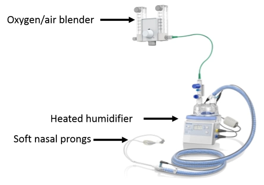
    1. Device (see picture above)
      1. Uses a special oxygen/air blender connected to a heated humidifier to saturate air with water and warm air to body temperature before delivery. This system allows for the delivery of very high flow rates (up to 60L/min)
      2. Connects to the nose with large soft prongs
        1. Potential benefits
          1. Ability to deliver heated and humidified gas at high flow rates
            1. Prevents drying of the airway and interference with mucocilliary clearance
            2. Enhances patient comfort
            3. May help moisten secretions making them easier to clear
            4. May lessen work of breathing by avoiding the bronchoconstricting effects of cold air and lessening the work needed to expectorate secretions
          2. Minimizing entrainment of room air
            1. Patients in respiratory failure often have high flow rates that exceed the flow of nasal cannulas and face-masks resulting in entrained room air which dilutes supplemental O2
            2. Flow rates in HFNC usually exceed patient-generated flows minimizing dilution of delivered oxygen
  • Improved ventilator efficiency
    1. HFNC can continually flush CO2 out of nasopharynx (lowering nasopharyngeal dead space) and allowing more of the minute ventilation to participate in gas exchange
    2. PEEP effect
      1. HFNC increases nasopharyngeal and esophageal pressure approximating levels seen with nasal CPAP
      2. HFNC may therefore provide a small amount of inspiratory assistance, help counter-balance auto-PEEP, and potentially improve oxygenation
    3. Breathing pattern
      1. Some evidence that HFNC helps increase TV and lowers RR
    4. Use
      1. Greatest benefit likely in patients with significant hypoxemia that would ordinarily be given standard high-flow oxygen therapy via a face mask
      2. Avoid in patients with high breathing workloads whose ventilatory failure is worsening (these patients require closely monitored NIV vs intubation)
      3. Usually provides more oxygen and gas flow than is necessary for patients with mild hypoxemia
      4. Initial settings
        1. Initial adjustments should be to flow rate as it is the flow rate that drives the physiologic benefit. Increase flow if RR fails to drop or if breathing remains labored with initial settings
        2. Starting flow rate is usually 35-40L/min
        3. Increasing flow rate should improve FiO2 as the amount of entrained air decreases
      5. There are two recent large randomized trials investigating the use of HFNC which are worth knowing
        1. High-flow oxygen through nasal cannula in acute hypoxemic respiratory failure (FLORALI) – NEJM, 2015
          1. Methods
            1. Multicenter randomized trial in 23 ICUs throughout France and Belgium
            2. Inclusion (need to meet all 4)
              1. RR>25, P/F < 300 while on O2 flow of 10L/min for at least 15 minutes, PaCO2<45, no history of chronic respiratory failure (ie pts with AHRF without concurrent hypercapnia)
            3. Exclusions
              1. PaCO2>45, exacerbation of asthma or chronic respiratory failure, cardiogenic pulmonary edema, neutropenia, shock, GCS <13, contraindications to NIV, urgent need for intubation, DNR (important to exclude obstructive lung disease and decompensated CHF where there is a known benefit to NIV)
            4. Patients randomized to 3 groups
              1. Standard O2 therapy: nonrebreather mask with flow of 10L/min or more with flow adjusted to maintain SpO2 >92% until patient recovered or was intubated
              2. HFNC: flow rate >50L/min and FiO2 100% with FiO2 adjusted to maintain SpO2 >92%. HFNC applied for 48 hours.  In the HFNC and standard oxygen group, a trial of NIV was allowed at the discretion of the MD
              3. NIV: pressure support adjusted to obtain a tidal volume of 7-10cc/kg PBW with initial PEEP between 2-10 cmH20. FiO2 and PEEP then adjusted to maintain SpO2 >92%.  Minimum duration of NIV was 8 hours/day for at least 2 calendar days.  NIV applied during sessions of at least 1 hour and could be resumed if RR was more than 25 and SpO2 <92%.  Between NIV, patients received HFNC
            5. Results
              1. 2,500 patients admitted with HRF  525 eligible  313 randomized (13% of patients with HRF included in trial)
              2. Causes of respiratory failure
                1. CAP: 64%
                2. HAP: 12%
              3. Initial mean settings
                1. Standard O2: O2 flow rate of 14L/min
                2. HFNC: O2 flow rate of 48L/min, mean FiO2 80%
                3. NIV: 8/5, FiO2 70%, TV 9 cc/kg
              4. 25% in NIV group received therapy for <4hrs per day
              5. 40 patients in the HFNC and standard O2 received BiPAP as a rescue therapy
              6. Outcomes (statistically significant results in bold)
HFNC NIV Standard O2 therapy
intubation rate by 28 days (primary outcome) 38% 50% 47%
intubation rate by 28 days in pts with P/F £200 35% 58% 53%
interval between enrollment and intubation 27° 27° 15°
ventilator-free days at day 28 24 19 22
ICU mortality 11% 25% 19%
90-day mortality 13% 31% 22%


  1. 90-day mortality
    1. Hazard ratio for death of 2.01 (1.01-3.99) when comparing standard O2 vs HFNC group and 2.5 (1.31-4.78) when comparing NIV vs HFNC
  2. Intensity of respiratory discomfort and dyspnea score significantly improved in the HFNC group 1 hour after enrollment
  • Conclusions
    1. The use of HFNC vs standard O2 or NIV did not prevent the primary outcome of need for intubation at 28 days
    2. HFNC was associated with several important secondary outcomes
      1. Lower ICU mortality
      2. Lower 90-day mortality
      3. Less need for intubation in patients with a P/F <200
    3. The authors and editorial wonder whether the high tidal volumes achieved with NIV may have contributed to the worse secondary outcomes seen in the NIV group
  1. High-Flow nasal oxygen vs noninvasive positive airway pressure in hypoxemic patients after cardiothoracic surgery – JAMA, 2015
    1. Respiratory failure is common following cardiac surgery with NIV currently the treatment of choice. However, NIV is cumbersome, requires significant resources, and fails in 20% of patients.   Authors wondered whether HFNC may be a better choice.
    2. Methods
      1. Multicenter randomized trial in 6 ICUs throughout France.
      2. Patients (any of the following)
        1. Failure of post-op SBT
        2. Successful SBT in patients with a BMI >30, LVEF <40%, or failure of prior extubations
        3. Successful SBT followed by failed extubation (P/F <300, RR >25 for at least 2hrs, use of accessory muscles)
        4. Exclusions included OSA, tracheostomy, DNI, delirium, nausea, vomiting, altered mental status, hemodynamic instability
      3. Patients randomized to the use of HFNC or NIV
  • Results
    1. 3,217 eligible, 830 randomized (80% were s/p CABG)
    2. Primary outcome of treatment failure (defined as re-intubation, switch to other study treatment, or premature study discontinuation)
      1. 9% in BiPAP vs 21.0% in HFNC (no difference)
      2. No difference in time to treatment failure (1 day in each group)
      3. Roughly 14% required re-intubation in each group (no difference)
      4. No difference in outcome among patients with a P/F < 200 (in contrast to the NEJM study).
      5. Similar dyspnea and comfort score
      6. No difference in ICU or 28 day mortality
    3. Conclusion
      1. HFNC is not inferior to NIV in preventing treatment failure in patients with or at risk for respiratory failure following cardiothoracic surgery


My thoughts

  • I find the results of the FLORALI study less convincing than the associated editorial suggests they are. The significant cross-over between groups (when not on NIV, patients in the NIV group received HFNC and 40 patients not on NIV were placed on NIV as a rescue therapy) and the variable duration of NIV therapy (25% of patients received therapy for <4 hours per day) make a clean comparison between the three O2 delivery methods difficult.  If a patient is on NIV for <4 hours a day and the other 20 hours was managed with HFNC, should their outcome really be associated with NIV use?
  • I also find it difficult to explain the improved 90-day mortality seen with HFNC.  It is hard to picture an interaction between any mode of non-invasive O2 delivery and mortality that does not hinge on precluding the need for mechanical ventilation which was not seen in this trial.  The authors of the trial and the editorial suggest that the high tidal volume achieved with NIV may have worsened lung injury thus leading to more time on the ventilator and increased mortality but it is hard to buy this conclusion if patients managed with NIV did not require intubation any more than patients in the other two groups nor did they have higher rates of refractory hypoxemia.  A trend of P/F ratios in all three arms over time would have been helpful.  Concerns have also been raised about the high rate of septic shock in the NIV arm (31%) vs the HFNC arm (18%)
  • Important to note that 45% of patients required intubation by 28 days. This highlights the importance of closely monitoring patients with AHRF being managed with HFNC or NIV as roughly half will fail. 
  • After reviewing this topic, I find the use of HFNC most appealing in patients who would normally be managed with a higher-flow face mask (venturi or nonrebreather). The ability to deliver very high flows of heated/humidified air seems to offer real physiologic and comfort benefits over typical higher flow devices.
  • We recently discussed the FLORALI study at a pulmonary conference. Faculty raised the important point that for older patients with pneumonia, difficulty with secretion clearance is often what leads to initiation of mechanical ventilation.  In this group of patients, placing them on NIV may inhibit their ability to clear secretions and hasten respiratory failure.  Specifically for this reason, Dr. Wunderink (MICU director) supported the idea of using HFNC over NIV as a first line therapy for older patients with pneumonia and resulting hypoxemic respiratory failure who do not require immediate mechanical ventilation.
  • For those who manage patients with hypoxemic respiratory failure (either in the ER or in the ICU), if you never find yourself initiating therapy with HFNC, you are probably underusing a helpful tool.


Take-home points

  • High flow nasal cannula devices deliver heated and humidified air at very high flow rates. Potential benefits include improved patient comfort, improved secretion clearance, a small PEEP effect, and washout of nasopharyngeal dead space
  • The use of HFNC may improve long-term outcomes in patients with acute hypoxemic respiratory failure although further trials are needed to validate this finding
  • HFNC is not inferior to NIV in preventing treatment failure following cardiothoracic surgery


Attached

  • Helpful review of HFNC from CHEST, 2015
  • FLORALI trial, NEJM, 2015
  • Randomized trial of HFNC following cardiothoracic surgery, JAMA, 2015


ILD Roundup 8/19/22

I. A patient with prior clinical diagnosis of pulmonary sarcoidosis presented to care at NMH with progressive pulmonary fibrosis. A transbronchial biopsy with EBUS was performed. The radiographic pattern and biopsy results were thought to be inconsistent with sarcoidosis; a differential of indeterminate UIP v NSIP (idiopathic v IPAF) remained. We discussed sending the patient for Envisia testing.


What do we mean when we say “Envisia”?

  • Envisia Genomic Classifier
    • Developed using machine learning applied to bulk RNA sequencing data from lung biopsy in combination with histologically-confirmed diagnoses
    • Helps differentiate UIP from non-UIP histologic patterns by transcriptomic signature

Envisia was validated in the BRAVE (Bronchial Sample Collection for a Novel Genomic Test) studies


How well can Envisia distinguish UIP from other pathology in conventional TBBx?

  • Envisia identified UIP in transbronchial biopsy with a specificity of 88% and sensitivity of 70%
  • Among patients who had “possible or inconsistent UIP” on HRCT, Envisia showed 81% positive predictive value for biopsy-proven UIP


Liu et al. BMJ 2022;377:e066354 https://doi.org/10.1136/bmj-2021-066354

Raghu et al. Lancet Respir Med 2019; 7: 487–96 10.1016/S2213-2600(19)30059-1


II. A patient who is undergoing evaluation for transplant had chronic hypersensitivity pneumonitis (cHP) in his differential diagnosis. We discussed the predictive value of BAL lymphocyte count given the fibrotic/chronic nature of his disease


What is the predictive value of BAL lymphocytosis in cHP?

  • A 2020 ERJ systematic review and meta-analysis suggested an association between BAL lymphocytosis and fibrotic cHP
    • BAL lymphocyte percentage higher in cHP (42.8%, CI 37.7-47.8) compared to IPF (10%, CI 6.9-13.1) and other IIPs
    • Analysis demonstrated that a BAL lymph% cutoff >20% optimized sensitivity and specificity for cHP compared to other IIPs

  • 2018 ATS/ERS/JRS/ALAT IPF diagnostic guidelines conditionally recommend BAL in patients with suspected IPF and a non-diagnostic HRCT pattern
    • A meta-analysis of eight studies in the most recent guidelines found no difference in BAL lymphocyte percentage between IPF and cHP

Morning Report 8/22/22 – Eosinophilic pleural effusion

Today’s morning report featured a fascinating case of eosinophilic pleural effusion from second year fellow Ted Cybulski

Second year PCCM fellow Ted Cybulski | http://tedc.cc/  | Twitter: @tdwck


First thing’s first – how do we define pleural fluid eosinophilia (PFE), and what are classic associations to be familiar with?


Some more quick facts:

  • Incidence estimated between 5-16% of all pleural effusions
  • More common in men (ratios reported between 2:1 and 9:1)
  • Malignant in roughly 35% of cases, of which 50% are lung ca.

Here a closer look at the broad categories on the differential diagnosis of PFE:


In our discussion, Dr. Sporn added that pleural fluid protein <4 and effusion size >1/3 hemithorax are suggestive against tuberculous effusion. A couple of other important points about TB effusion:

  • Can occur with primary or reactivation of infx
  • Pleural fluid ADA >40 u/L argue strongly for TB especially in lymphocytic exudative effusions (90% of cases)
  • Eosinophilic effusions are relatively rarer presentation

Below Ted details the association of eosinophil count in PFE with malignancy – while a lower count (<40%) is MC in malignancy, a higher count is not necessarily reassuring. Other characteristics which carry higher risk for malignant effusion include advanced age and higher pleural fluid LDH (cutoff >900 suggested)


Takeaways

  • Pleural fluid eosinophilia = >10% eos, found in 6-14% of pleural effusions
  • Classic association with trauma, repeated taps, asbestos.
  • MC etio malignant
  • Lower eos (10-40%), higher LDH (>900) and advanced age associated with higher probability of malignancy
  • TB can present with eosinophilic effusion, but classic presentation is lymphocytic exudate with high ADA (>40)

Thanks for leading a great discussion, Ted!

Sources cited

  • Krenke et al. ERJ 2009; 34(5):1111-1117 (https://pubmed.ncbi.nlm.nih.gov/19386682/)
  • JM Porcel. Lung 2009; 187:263-70 (https://pubmed.ncbi.nlm.nih.gov/19672657/)

“Effect of postextubation high-flow nasal cannula vs conventional oxygen therapy on reintubation in low-risk patients,” JAMA, 2016, Spain

“Effect of postextubation high-flow nasal cannula vs conventional oxygen therapy on reintubation in low-risk patients,” JAMA, 2016, Spain

Question: Does HFNC reduce the need for reintubation in patients at low risk of post-extubation respiratory failure?

Study Type: Multicenter, randomized clinical trial in 7 ICUs in Spain

Study Population: Patients who passed an SBT after at least 12 hrs of mechanical ventilation were eligible if they meet the following inclusion criteria: Age <65, not initially intubated for CHF, absence of mod-severe COPD, APACHE II <12, BMI <30, no known airway problems and low risk of developing laryngeal edema, adequate cough and requiring suctioning <2xs Q8hrs, not difficult to wean, mechanical ventilation <7 days, <2 co-morbidities.  Patients were excluded if they had a tracheostomy or had evidence of hypercapnia during an SBT.

Study Groups: Patients in the intervention arm were placed on HFNC with flow set at 10L/min which was titrated up at 5L/min intervals until pts experienced discomfort.  FIO2 was titrated to keep SpO2 > 92%.  Patients in the control arm had conventional oxygen applied through a facemask or nasal cannula titrated to keep SpO2>92% for 24 hrs.

Primary Outcome: Need for reintubation at 72 hrs.

 Results: 527 patients randomized. Notable patient characteristics: primary neurologic diagnosis (29%), scheduled or urgent surgery at admission (47%), primary respiratory failure (17%).  HFNC significantly reduced the need for reintubation at 72 hours (4.9% vs 12.2%, p=0.004) with a number needed to treat to prevent one reintubation of 14.  Patients treated with HFNC also had lower rates of reintubation secondary to respiratory causes (8.3% vs 14.4%, p=0.03). There was no difference in ICU length of stay or mortality.

Caveats: Primarily neurology or surgery patients (not a typical MICU population), most common cause for reintubation in the control arm was inability to clear secretions which is perhaps a function of having so many pts with neurologic injuries.

 Take-home Point: In a cohort of mostly surgical and neurologic patients at low risk for reintubation, HFNC reduced the need for reintubation compared to conventional oxygen therapy.  It is unclear if these results are generalizable to a more typical MICU population.


ILD roundup 8/12/22

I. A 62 yo female with family history of pulmonary fibrosis with an NSIP pattern

What is the relationship between telomere length and familial pulmonary fibrosis?

-Heterozygous mutations in coding regions of two telomerase genes (TERT :: protein component, TERC :: RNA component) have been associated (found in 15% of kindreds) with familial IPF  

Pulmonary fibrosis is seen in 50% of women and 60% of men 60 years and older with TERT mutations


-UIP radiograph pattern (74% HRCT) and UIP pathologic pattern (86% surgical lung biopsy) is most commonly encountered 

-Even in sporadic cases of IPF, a significantly higher proportion of individuals have telomere lengths <10th percentile compared with controls, even absent TERT/TERC mutations (present in 1-3% of sporadic cases) 


Plotting mean telomere length against age with 10-90th percentile in normal controls mapped in blue region


https://www.atsjournals.org/doi/pdf/10.1164/rccm.200804-550OC 

https://doi.org/10.1371/journal.pone.0010680 

II. 69 yo F with seropositive RA and progressive fibrosing ILD, found to have upper zone predominant fibrosis, honeycombing, TBE.  

What HRCT & histopathologic patterns are most commonly associated with RA-ILD?
 -UIP pattern on HRCT most commonly encountered, between 40-60% of cases.  

-Histopathology usually correlates with HRCT. MC encountered histopathologic patterns are UIP, NSIP, OP 


Aggregated data from multiple studies demonstrating the most common histopathologic patterns in CTD-ILDs. Note larger proportion of UIP in RA-ILD


-RA-ILD associated with UIP pattern has an increased risk of disease progression and death, and increased risk of flares compared with RA-ILD with NSIP pattern 

Kaplan-Meier curve showing RA-ILD with UIP pattern; cumulative survival intermediate between RA-ILD with NSIP and IPF (another ILD characterized by UIP HRCT and histopath)


What is the role of antifibrotic therapy in non-IPF ILD? 

– A recent trial (INBUILD – 2019) looked at nintedanib (antifibrotic FDA approved for IPF) in patients with non-IPF fibrosing interstitial lung disease (n=663) over 12 months of follow-up 


Figure demonstrating the primary endpoint, decline in FVC over period of followup (188 mL in placebo versus 80 mL in nintedanib group).


-As a secondary endpoint, mortality was less in intervention group (11.5% placebo vs  8.1% nintedanib) although this was not statistically significant 

– Since publication of INBUILD trial, FDA has approved use of nintedanib/Ofev for non-IPF progressive fibrosing ILDs 

Finally, for a current and comprehensive review of the treatment of fibrosing interstitial lung disease including IPF and other progressive pulmonary fibrosis (PPF), make sure to check out a recent review in BMJ by our very own Gabby Liu, Scott Budinger and Jane Dematte!  

An algorithmic approach to diagnosis and management


https://doi.org/10.1378/chest.09-0444 

https://www.nejm.org/doi/full/10.1056/NEJMoa1908681 

https://www.bmj.com/content/377/bmj-2021-066354 

https://www.fda.gov/news-events/press-announcements/fda-approves-first-treatment-group-progressive-interstitial-lung-diseases 


HHT Review

Callback to this morning report.


Grand Rounds 8/19/22 – Anti-MDA5 ILD

Thanks, Tom Bolig, for presenting on idiopathic inflammatory myopathy-associated ILD, focusing on anti-MDA5 ILD.

My takeaways:

  • while a dermatomyositis, there are mild to no muscle symptoms; there are hallmark cutaneous manifestations though
  • is important to test for specific antibodies and involve Rheumatology early
  • lower overall survival and higher risk for developing a rapidly-progressive ILD
  • may be triggered by anti-TNFalpha therapy; some mimicking of psoriatic arthritis
  • more specific cutaneous phenotypes are skin ulcerations (OR 18) and palmar papules
  • no guideline-recommended treatment but common to see combination immunosuppression such as glucocorticoid with calcineurin inhibitors (tacrolimus)
  • there may be benefit to triple therapy up front compared with stepwise
  • salve therapy with plasma exchange is promising, as are use of rituximab and tofacitnib

Thanks, Tom!