Student Blog

Finding Neverland in the Netherlands: My Summer in an Art Heritage and Science Wonderland

Clockwise from top left: about to land at Schipol Airport; a scene taken during my daily bike commute to the lab; a private demonstration at De Kat, the only windmill still producing pigment; the Rijksmuseum and IAmsterdam sign

“There are some things you simply cannot prepare for,” I thought as the plane began the final descent over my home for the summer, the Netherlands. This first brush left me in awe– anxious to uncover the layers that make up this country’s unique character and rich past.

I had no idea that bike “traffic” existed, much less that there is a rush-hour for such traffic. I could not have anticipated being inches away from real-life actual Rembrandt paintings on our very first day. It is overwhelming to be in the presence of such revered masterpieces, and to contemplate how the artists of the Dutch Golden Age could achieve such strikingly realistic works. Even after seeing Girl with a Pearl Earring in person, I’m not sure I’ll ever understand how Vermeer was able to capture light in a bottle and apply it to canvas. Being in the center of this art hotspot feels like the luckiest day ever: as if you’d stumbled upon a whole field of four-leaf clovers, bathed overhead by a quadruple rainbow. Only in the Netherlands could I experience so much of the Dutch Golden Age. There have been so many once-in-a-lifetime experiences as I’ve learned about art conservation and Dutch cultural heritage.

The power of science to preserve these pieces is exhilarating. Admittedly, my background is in materials science and engineering, and so it has been eye-opening to be introduced to the art conservation field and its scientific, historical, cultural, and ethical components. I’ve learned it is a far cry from an easy task to preserve art; the endeavor seems quite chaotic when you stop to think of all the variables. What is the paint composition? What is the chemistry of the binders? How thick is each paint layer, and was it allowed to dry before the next layer was painted? What would the painting have looked like at the time of creation? This is just a tiny sample of questions that art conservators, art historians, and scientists must answer together.

As a student in the NU-IRES program, I conduct research at the University of Amsterdam (UvA) and make my own contribution to the art conservation community. My undergraduate research at Rutgers University heavily focused on characterization of polymer composites, and I became particularly interested in rheology. I am fortunate to continue studying rheology at Northwestern University as a graduate student in the Shull Group. Our group works extensively with the quartz crystal microbalance (QCM) to study polymer thin films. QCM is a powerful technique and—depending on film thickness– can be sensitive to mass changes and viscoelastic properties. At Northwestern, I have been using the QCM to investigate the effect of molecular weight on glass transition temperature in polystyrene thin films. A great advantage of the QCM is its portability. After my last final of the academic year, I packed the QCM equipment in a small cereal-sized box and took the lab on the road.

After landing in one of the art capitals of the world, it was time to shift to a material that art conservators and scientists really care about: linseed oil. I am investigating the QCM’s potential to study the early curing stages of this drying oil. My Dutch advisor, Dr. Piet Iedema, and his group have developed computational models to describe the mechanisms occurring in these early stages. I’m interested in developing a QCM experiment to study the changes in mass when a raw linseed oil film is applied to the QCM crystal. Linseed oil is composed of a hodgepodge of fatty acids; namely -linolenic, oleic, and linoleic acid. Atmospheric oxygen molecules react with the unsaturated bonds in these fatty acids and create highly reactive peroxide species, which then initiate a whole cascade of reactions. During these processes, the oxygen absorption corresponds to an increase in mass. The linseed oil also hardens during the cure process. I’m interested in using the QCM to monitor changes in both mass and viscoelasticity. Linseed oil has been more difficult than polystyrene to work with, and my equipment started malfunctioning. Fortunately, my best friend had planned to come visit in mid-July and was able to come with some replacement equipment from the Shull lab. But even with that stroke of luck, there’s always a risk of persistent problems. These various challenges provide great learning opportunities. So while I may not have been able to conduct as many experiments with linseed oil as I had hoped– I am still learning valuable information about QCM troubleshooting, circuits, mechanics, paintings, metal soaps, etc.

I am immensely grateful for the support of Dr. Shull, Dr. Iedema, Dr. Walton, and Dr. Gambardella this summer. Dr. Shull’s advising and troubleshooting help via email and weekly Skype calls has been a real lifeline. I thank him for his encouragement to apply for the NU-IRES program. The time here has been transformative in ways I could not have anticipated, but sometimes it’s fantastic to have your breath taken away.

Art and Science: The Amsterdam Experience

Posing in front of the Rijksmuseum in the famous “I Amsterdam” sign.

My name is Francesca Long and I am just entering into the second year of my PhD in materials science at Northwestern University. I am lucky enough to spend two months this summer in Amsterdam as part of the Cultural Heritage in the Netherlands program: a National Science Foundation (NSF) sponsored International Research Experience for Students (IRES). My undergraduate degree is in Materials Science and I focused in metallurgy, the topic of my graduate research as well. Back in the US, I study cobalt-based super alloys and do a lot of mechanical testing and microscopy to study my samples. Despite this background in metals, I had always had a strong passion and interest in the arts. I did a minor in Classical Studies alongside my bachelor’s degree and was able to take a class that blended art and Materials Science both in my senior year of undergrad and my first year of graduate school. The course I took in graduate school was all about the field of art conservation from the materials science point of view and really opened my eyes to this field. It became quite clear that this was something I was truly passionate about and wanted to be able to be a part of.

My favorite place to eat my lunch: in the Rijksmuseum garden.

I was fortunate enough to have been allowed to come to Amsterdam to get my first real experience in this field of research at the Ateliergebouw Rijksmuseum. My project is focused on the degradation of arsenic sulfide-based pigments. Artists use many layers in their works to achieve their desired final look, however over time the surface can change in appearance. It is well known that arsenic sulfide pigments, specifically Orpiment and Realgar, are very sensitive to light and moisture in the air and within the paint layers and as such they break down over time and can lose their gorgeous yellow and orange hues. I will be using scanning electron microscopy (SEM) and energy-dispersive x-ray spectroscopy (EDX) to analyze cross sections thought or known to contain these pigments in order to determine: the presence of orpiment and/or realgar and the composition of the paint layer as a whole. By knowing what else is present, we hope to add further insight into how these rich pigments go through such a dramatic change and lose their vibrant color and turn white or transparent with age. In addition to this, I am working in collaboration with a conservator who is translating paint recipes in order to hopefully pair up the paints we study in cross sections with old treatises to know exactly what recipes an artist used.

The Netherlands has a very strong national pride in their art. Rembrandt’s Night Watch is beloved all around the world but especially here among countless others. It is this dedication as a whole towards preserving their cultural icons that makes the Netherlands the perfect place for this kind of research. It is something people are fiercely passionate about and as such the resources needed to study how best to preserve and protect works of art are readily available. Not only that, but being in the context of the paintings one is studying makes all the difference in the world. I can look at a cross section and see on and online database what painting it came from and where, but to be able to walk across the street into the Rijksmuseum and see that same painting on the wall in it’s full glory is indescribable. It puts the work into perspective and makes it seem all that much more important.

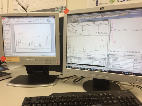
EDX elemental mapping (right monitor) and corresponding SEM image (left monitor) of a cross section from a De Heem still life on a day it was cooperating with me.

I think doing research abroad has so many benefits to simply staying in the US and working with collaborators in different countries. It exposes one to new languages and cultures and allows one to immerse themselves in their work while experiencing all that another country has to offer. The relationships I can form and build on while here will carry on long after I leave the US and open up the doors in terms of future collaborations. It removes the distractions of being at home and allows the science to become the main focus of the summer. This is a wonderful opportunity and one I would highly recommend to anyone with even the slightest thought of wanting to do a program like this.

As with any research project, I quickly learned that things don’t always go to plan immediately. Equipment can break and take time to get working again which is always frustrating. However, when the SEM computer started giving up, it allowed me to attend a conference that was going on at the Ateliergebouw where I heard a lot of interesting talks about different techniques being developed for use by conservators to study their works safely. I have learned more about artistic methods and painting style in the last three weeks than I ever thought possible. I have learned that I really do want to continue working in this field because every day I am excited to go into the lab and do experiments or even to just sit at my desk and read papers about this line of work. I have also learned that the Dutch are far better at speaking English than I am at speaking Dutch (which isn’t saying much as I have only mastered a few words in Dutch so far) and are so welcoming and happy to help and share their own research with me. I never felt like my lack of experience in this field was a hindrance, it only meant that there was that much more for me to learn every day that I am here and for me to continue learning over the weeks to come.

As Interesting As Watching Paint Dry: The Analysis of Twentieth Century Oil Paints

The Er:YAG laser

My name is Samantha Miller, and I am a Colorado State University graduate student researching in Amsterdam as a part of the Cultural Heritage Science Research funded by the National Science Foundation. As a participant in this International Research Experience for Students (IRES), my work is focused on the chemistry of art conservation. I particularly enjoy this field because it brings together both art and science. By unifying the two, this work defies the stereotypical expectation that the two must be in constant rivalry in order to flourish. My Dutch mentor, Dr. Klaas Jan van der Berg, is a senior conservation scientist and has worked closely with me as I apply my background in analytical chemistry to the analysis of twentieth century oil paints.

Although the old adage “as interesting as watching paint dry” sarcastically illustrates the process as passive and mundane, a number of processes are occurring at the molecular level which alter the chemical composition of the paints. Common drying oils such as linseed and safflower oil are mixed with artists’ pigments and later undergo a series of autoxidation reactions in which triacylglycerides form a crosslinked polymer network as the paint dries. The presence of tri-, di-, and mono- glycerides can be monitored using Mass Spectrometry, a powerful technique that separates ions based on their mass-to-charge ratio. By identifying the presence of these different fatty acids, along with other key compounds such as palmitic acid, stearate, linoleic acid, oleic acid, etc., it’s possible to construct some preliminary commentary on the age of the oil paint as well as the conditions in which it aged.

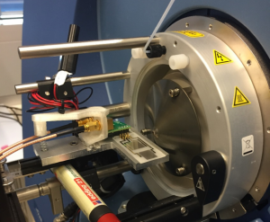
The quadrupole mass spectrometer with the SAWN chip accessory

My favorite part of chemistry is working with the instrumentation. Obtaining the spectra of different oil paints imparts a more poetic end to the passive drying process. By the end of my tenure here in Amsterdam, I hope to demonstrate that reproducible data can be obtained not only using electrospray ionization (ESI) mass spectrometry, but surface acoustic wave nebulization (SAWN) mass spectrometry as well. Just as the name implies, electrospray techniques use differential voltages across directing cones to generate a gentle stream of ions that pass into mass spectrometer, in our case, a quadruople time-of-flight spectrometer. SAWN uses an applied voltage to generate a standing wave whose energy is transferred to the analyte. Once a threshold energy is reached, the analyte is nebulized into a plume which directed into the mass spectrometer. Preliminary tests have shown that SAWN requires much less volume and adds the additional benefit of removing the ever temperamental injection equipment of ESI methods. Eliminating the pumps, capillary tubes, and injection syringes not only lowers the possibility that a faulty part will slow the experiment, but also the probability of introducing contaminants. The techniques differ only in the way the oil paint is ionized, and if SAWN proves to be a successful method, then hopefully the Rijksmuseum will be able to apply this softer and quicker ionization method to future oil paint samples as well.

As glamorous and photogenic as the research experience has been, the most enjoyable part about working at the Rijksmuseum and Amsterdam is collaborating with fellow scientists. Walking through the main hall of the Ateliergebouw or the University Science Park, it’s easy to see that everyone is absorbed in their work. Yet, beneath the lab coats or behind the monitors are people who share a common passion for both art and science. The scientific community here in Amsterdam has a tangible awareness that they are working towards something greater than their own conservation project or publication. Each office and laboratory I pass reminds me of a different gallery in an art museum; Each is littered with vestiges of their specialized field: spectroscopy, spectrometry, rheology, proteomics. Everyone represents a different facet of science through their approach to conservation. It’s humbling to think that together, the individual efforts, failures, and successes somehow combine to let national artifacts live on for a new audience to appreciate and treasure the way we have.

Sitting at the Intersection between Art and Medicine

My name is Gabriela Diaz and I am a 3rd year student at Texas A&M University, Kingsville pursuing a Bachelor of Science in Chemical Engineering. My background in cultural heritage is a research experience (NSF-REU) at the University of North Texas studying X-ray fluorescence (XRF) to characterization 19th and 20th century silver-plated objects from the Dallas Museum of Art. I am here in Amsterdam through the Cultural Heritage Research in the Netherlands, an International Research Experience for Students (IRES) which is sponsored by the National Science Foundation (NSF).
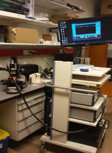
The focus of my summer research is a technique used primarily in the medical sciences but one that is rapidly gaining popularity in the art conservation community: optical coherence tomography (OCT).  This reseach brings me to work most often in the Academic Medical Center under the mentorship of forensic biophysicist Dr. Maurice Aalders.  However , since our primary aim is to use OCT at the Rijksmuseum, I also work with Katrien Keune in their atelier building. The technique operates on semi-transparent surfaces and is most famously used to understand the structure of the eye but has recently been applied in paintings conservation.

My research question explores how light penetrates the surface and stratigraphy of painting varnish layers. The chosen varnishes I will be working with will be dammar and mastic resin. Using OCT we measure the refractive index of different varnish recipes and also visualize how they  appear within layering scenarios. We can also explore which situations give false positives and false negatives in order to take some of the guess work out of OCT analysis.

The Netherlands holds a special place in my heart due to the fact that one of my favorite painters is Johannes Vermeer. One of the theories behind his success is his prodigious use of optics—adapting the camera obscura—in order to “paint with light.” It is certainly a wonderful coincidence that I, too, shall be operating lenses and mirrors to tell a story—though my case is through an interferometer inside the OCT. Studying these methods of art conservation abroad yields the opportunity to gain new perspective on the styles and approaches scientists outside the U.S. take to restore a work of art. Being in the Netherlands also gives me a chance to get to know a bit about the culture of these world-renowned masters as well as admire their beautiful country.

So far I have learned that no matter how well you think you understand something there will always be something unexpected to bring you back to the scientific chase. I never imagined finding myself studying methods to analyze art inside a hospital, and yet as the days pass it makes perfect sense that skills applied in preserving the health of an individual can find some application in preserving the health of a great painting. Everyday brings new questions and new things to image under the scan-head of the OCT. This constant curious thrill as well as the immersion in the energy of the city has made my transition from the states an adventure to remember.
Rembrandt and Surface-Enhanced Raman Spectroscopy (SERS)

In the lab with the Renishaw inVia Raman Microscope at the Ateliergebouw Rijksmuseum

My name is Shelle Butler and I am a chemistry graduate student from the College of William and Mary conducting summer research for cultural heritage in the Netherlands with the International Research Experience for Students (IRES) program funded by the National Science Foundation (NSF). Back home in Williamsburg, Virginia under the mentorship of Dr. Kristin Wustholz and Colonial Williamsburg Foundation senior paintings conservator Shelley Svoboda, I perform surface-enhanced Raman spectroscopy (SERS) analysis on microscopic samples and cross sections from paintings to identify pigments and binding media.

Now, I find myself in a new land, the Netherlands. This once in a lifetime collaboration transports me from the early American colonial period to the Dutch Golden Age, where Rembrandt Harmenszoon van Rijn and Johannes Vermeer painted their masterpieces of Dutch history and the intimate scenes of Dutch domestic life in the 17th century. I bring with me an innate curiosity, a passion for both art and science, and my SERS experience.

Taking in the beauty of Dutch culture by bicycle

So, why is SERS so important? It can often be difficult to identify a specific pigment by other analysis techniques. The use of light microscopy and scanning electron microscopy along with X-ray elemental analyses can be used to elucidate elemental composition in the paint cross-section, but does not identify some pigments, called lakes, whose color is not derived from minerals but from organic colorants.. Additionally, high-performance liquid chromatography (HPLC) is commonly used to identify the organic dye of the pigment, but requires a larger sample. When we are talking about precious artworks, we want a minimally to non-destructive analytical technique.

Raman spectroscopy is a vibrational spectroscopic technique commonly applied to reveal a structural fingerprint, which can be utilized to identify molecules. Normal Raman spectroscopy works well with many pigment samples, such as the strong signal observed with vermillion, a red inorganic pigment from the mineral cinnabar. However, for some samples, like red organic lake pigments, the Raman signal is too weak and is often hidden by powerful fluorescence, which conceals the chemical fingerprint needed for identification. To reveal the hidden information, the SERS technique is applied by adding nanoparticles made from noble metals (e.g. silver, gold, or platinum) to the surface of the sample to serve as antennae that amplify the Raman signal and subdue interference caused by fluorescence.

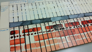
Madder lake and lead white references curtesy of the Rijksmuseum

For paintings conservation, the SERS technique can be used to answer questions about the artist’s palette, their original intent, and can aid in digital reconstruction to visualize the painting’s original appearance to virtually resolve centuries of fading and degradation caused by light exposure and other environmental factors. Together with Dutch scientists at the Rijksmuseum, I am working to develop a Raman database and a protocol for SERS cross section analysis for red lake pigments, including carmine lake (a pigment made from the cochineal insect), madder lake (a pigment made from plant root), and brazilwood (from the tropical hardwood). I am starting with known reference material for protocol development and optimization. Once this has been accomplished, we can then move on to the analysis of works by the Dutch Golden Age master painter, Rembrandt!

A Vivid Summer in Amsterdam

katherineMy name is Katherine Ferreras and I am a senior at The City College of New York (CCNY) of The City University of New York, pursuing a Bachelor of Science in Environmental Chemistry, and a minor in Earth and Atmospheric Science. This summer I am in Amsterdam as part of Cultural Heritage Research in the Netherlands, a NSF-funded International Research Experience for US Students (IRES) in the Netherlands. Under the mentorship of Dr. Urs Jans and close collaboration of Dr. Glen Kowach at my home institution, I have been able to explore and expand my interest in groundwater remediation techniques. My work focuses on the development of green rust, a highly reactive iron (II) mineral capable of reducing a range of inorganic and organic species. As this includes toxic materials and organic pollutants, this project can help to provide the necessary knowledge for a new approach to remediate contaminated aquifers. The motivation of my project is to understand the structure, behavior and reductive kinetics of the reaction between green rust and chlorinated hydrocarbons, since this is a possible effective approach to reduce nonpolar chemicals, such as carbon tetrachloride that are found in ponds at the bottom of aquifers.
asd
Interpretation of UHPLC – DPA detection analysis.

In search of exposure to a different application of chemistry, my mentors encouraged me to apply to this program. I have discovered that cultural heritage science is a multidisciplinary field with unique and interesting challenges. At the Rijksmuseum, under the guidance of Dr. Maarten van Bommel, my project focuses on the development of an ultra high performance liquid chromatography (UHPLC) method for the separation of natural, acid, and basic synthetic dyes. Dyes are present in a range of objects, including textiles, paintings, and wooden objects. Identification of dyes from cultural heritage objects is essential to help conservators create an adequate restoration approach. Currently, dye identification requires several different tests: development of a single chromatographic technique for these early dyes would prevent excessive destructive sampling of these precious cultural objects.

Picture1
Enjoying the beach at Den Haag, The Netherlands’ third-largest town

The Netherlands has done a wonderful job of merging university and museum research, so it has been an ideal place to experience a new setting for research outside of strict university settings. This program has given me the opportunity of listening and learning from several talks presented by Ph.D. candidates, scientists, and conservators at such an early stage of my education. Topics as the degradation of ultramarine, the formation of metal-soaps in paints, and the selection of solvents to restore cultural heritage are not longer completely unfamiliar for me. Thanks to the great support and guidance of Dr. Maarten van Bommel I have gotten to clearly understand the science and instrumentation behind UHPLC.

I applied to this program seeking new experiences, not just in the science field but also in a new environment. Being my first experience abroad, I can say that the architecture, the wide selection of art, and the cycling around the city along the canals have made the transition from my comfort zone to an overseas adventure very enjoyable.

A Scientific Summer in Amsterdam!

PictuqWDre1
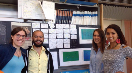
Some fabulous members of the Chromatography group at UvA (From left: Fleur, me, Charlotte, and Serafine)

My name is Abed Haddad and I am currently in Amsterdam as part of the Cultural Heritage Research In the Netherlands an International Research Experience for Students (IRES) program sponsored by the National Science Foundation (NSF). In my undergraduate studies, I delved into both chemistry and art history with passion.  Unbeknownst to me, there was a whole field that combines both! And now, I am a graduate student at the City University of New York under the mentorship of Dr. John Lombardi who is also a member of the Scientific Research Committee at the Metropolitan Museum of Art. My work in Surface Enhanced Raman Spectroscopy (SERS) lies at the intersection of nanotechnology, material chemistry, and forensic sciences, and has become of great interest to conservators because of its noninvasive nature. SERS uses the interaction between lasers and nanoparticles to identify colorant at very small concentrations due to physical and chemical enhancement mechanisms. As an ultra-sensitive analytical technique, it has been successfully implemented for cultural heritage research at the Metropolitan Museum; for example, the identification of dyes used in the Unicorn Tapestries at the Cloister, or early synthetic dyes used in late 19th century Japanese prints.

effff
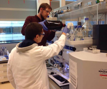
Here, Bob and Reuben are setting up the LC instrument at UvA for a Size Exclusion study of polymers

Still, it is often difficult to sum up the work of cultural heritage scientists; and that’s one of the exciting parts of the job. We wear many hats as we collaborate with art historians, conservators, restorers, and other scientists to uncover truths about our history, and I hope to do just that during my time in the Netherlands!

In partnership with the Rijksmuseum and the University of Amsterdam (UvA), my project exploits Ultra-High Performance Liquid Chromatography (UPLC) for the study of natural and synthetic textile dyes. UPLC utilizes size or overall charge characteristics to separate molecules as they are forced with high pressure through a separation column packed with surface-modified silica beads. Investigating comprehensive analysis parameters for rapid and effective separation of complex mixtures is of immense forensic value. Two different separation factors can be compounded for a 2-dimensional evaluation, increasing the identification capacity of an LC system, as dyers would sometimes use more than ten different colorants to achieve a particular shade. Furthermore, we hope to directly analyze the eluted compounds with mass spectrometry to establish degradation products and the mechanisms by which they occur. A well-resolved separation allows conservators to understand how colorants fade over time.

wRG
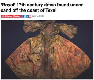
News article discussing the newly discovered garment

The cultural treasures of the Netherlands, combined with the sizeable cohort of conservation professionals at the Rijksmuseum, make for incredible study opportunities. Our research will be applied to an exemplary Dutch discovery from this year: a luxurious and rare 17th century dress found by divers off the coast of Texel amongst other small objects. The fabric is in dire shape, which requires diligent examination of small sample sizes. Alongside Professor Maarten Van Bommel and Ph.D. candidate Bob Pirok, we hope to undertake a substantial study of dyes used and uncover much about the provenance of the wardrobe.

I am relishing my time in beautiful Amsterdam by getting to know its streets and people; after all, not all of my time will be spent in the lab. I am looking forward to volunteering at Queeristan, an autonomous festival for radical queer politics, especially in the testing times following the Orlando shooting. Plus, the Netherlands has much to offer outside the capital. I am particularly excited to visit the Rietveld Schröder House in Utrecht, the only building truly designed after the abstract tenants of De Stijl.

Truly, I hope to inspire other scientists to make their own contribution to preserving the records of human creativity and ingenuity, and I cannot express how fortunate I feel to be part of this remarkable program.


Proost!


When Color is Sensitive to Light

Marcie1
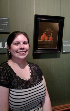
With Still Life with Five Apricots by Adriaen Coorte, a painting with migrating arsenic

My name is Marcie Wiggins and I am participating in the NSF-funded International Research Experience for US Students (IRES) this summer in the Netherlands. I have been interested in conservation and cultural heritage studies since high school as a unique, challenging way to utilize chemistry. Pursuing chemistry and art history as an undergraduate at the University of Maryland, I was able to further explore this interest through internships at the Library of Congress and the Smithsonian’s Museum Conservation Institute. Now, I continue this focus as an analytical graduate student at the University of Delaware. Through my experiences in cultural heritage science, I have seen the various ways cultural heritage studies have benefited through partnerships with the sciences.

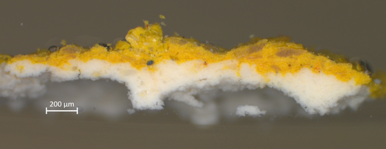
Paintings provide a challenging chemical system for many reasons, one of which is the multiple layers of paint an artist can use in compositions. As a result, below the visible surface there can be many distinctive layers interacting and affecting the object. My summer research, mentored by Dr. Katrien Keune, will be addressing a recent phenomenon concerning arsenic paints, such as orpiment (As2S3), where arsenic degradation products have been found throughout other layers in a painting, from wood support to the top varnish layer. These pigments are sensitive to light, which accelerates degradation. As arsenic sulfide pigments photo-degrade, they turn into arsenic trioxide, which is slightly soluble. As a result, it is believed the arsenic is transported throughout the paint layers with water, where the arsenic may react with other compounds as well. This can pose a risk for conservators interested in removing the old varnish layers and can influence how they handle the object’s treatment, as this can causes bright oranges to loose some of their color.

marcie2
A cross-section of orpiment paint over a lead white ground prior to degradation

To study the transportation of arsenic species as a function of relative humidity, model samples of orpiment on differing grounds will be exposed to a range of relative humidities in open and closed systems. These types of weathering experiments are common in cultural heritage research to reconstruct objects degradations to better prevent the processes. This is similar how car coatings are put through extreme conditions to improve them. Cross-section samples will be taken during the process allowing us to study the multilayer system. This means samples will be embedded in resin, turned on their side, and polished to expose all the layers on the paint sample. Over these past two weeks, I have been setting up humidity chambers using salt solutions and monitoring the systems. I am now focusing on making cross sections of the controls and initial samples, which will next be analyzed with light microscope, mapping-FT infrared and mapping Raman spectroscopy, X-Ray diffraction, and Scanning electron microscopy with elemental analysis.

marcie3
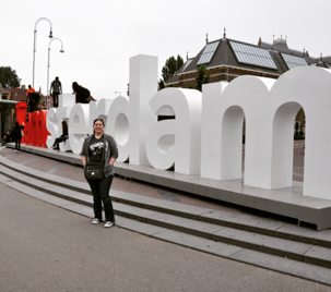
The iconic “I Amsterdam” sign outside of the Rijksmuseum

Working at the Rijksmuseum in Amsterdam in an effort to tackle these cultural heritage questions is a dream come true! During my art history classes, many fantastic paintings were housed in the Netherlands, so it will be great to see those same paintings in person finally. But more importantly, it is exciting to work alongside conservators and scientists on such a vast collection of cultural heritage objects. Already I have gotten to not only work with historical cross-sections, but I have gotten an up close look at these objects in the conservation labs, instead of just being sent cross-sections to analyze from across the world. A conservator, Nouchka de Keyser, showed me a Jan Davidsz. de Heem painting containing arsenic paint to analyze alongside my reconstructions, which has helped put the whole project into perspective. Having worked in and with museums in the US for several years, I have wanted to broaden my experiences beyond the US. I am looking forward to learning the different approaches and techniques abroad to return to Delaware with. I hope to forge collaborations and partnerships that will last beyond this summer, as I have already found this a great place to work and a lovely country.

Paintings, Not as “Still Life” as you might think

MyPicture1 name is Lindsay Oakley and I am a Northwestern University graduate student working in the Netherlands this summer as part of a NSF sponsored International Research Experience for Students (IRES) focused on investigating questions in cultural heritage science.  I was first introduced to the field as an undergraduate at the College of William and Mary. When I was struggling to settle into a major, deciding between chemistry and history classes that I also enjoyed, I met a chemistry professor setting up a collaboration with the paintings conservator for Colonial Williamsburg.  I volunteered to get involved and was quickly fascinated by the way that the scientific study of objects can reveal insights into people and technologies of the past. But historical knowledge also serves to help interpret scientific results, and this synergy leads to new understanding.  This experience led me to Northwestern and a project in partnership with the Art Institute of Chicago where I have continued studying paint and new tools that can be applied to understand it and preserve it for future generations.
Many perceive paintings as static objects, capturing an image from an artist’s mind in perpetuity.  In reality, from the time an artist touched a brush to canvas until you encounter the work 10s or even 100s of years later, quite a lot has changed. The colors may have faded, a wrinkling or cracking pattern may have developed or new chemical species that are products of aging and degradation reactions may have formed and started to disturb the surface of the painting.  (For an interesting article on this last example, you can read more here.) But how do these things happen?  What physical or chemical processes are responsible?  These can be difficult to observe and quantify.  Sometimes the processes are just too slow (at least for one PhD student!) and sometimes we need to study the mechanisms of interest at the molecular level.  In these types of dilemmas, we can turn to computer models for help.

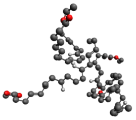
If you zoomed in on a painting down to the molecular level, this is a representation of what you would find. Long chain molecules that make up the paint binder link together to form a network that small molecules can diffuse through.

This is why I am so excited to be here in Amsterdam this summer.  Over the course of the next seven weeks I will be building a computer model of an oil paint layer and exploring how small molecules move and diffuse through it, while working with some of the leading experts in modeling and paint dynamics. For aging and degradation reactions to occur, reacting species have to “find” each other in the paint layers. With experiments we can observe where these species end up, and this study will seek to answer complementary fundamental questions about how they are transported.  We will model the oil paint as a field of atoms that exert forces on the molecules of interest, either attractive or repulsive.  Setting up this field will be the main research challenge I will face in the coming weeks.

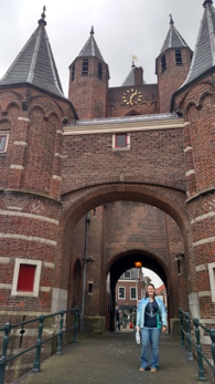
At the Amsterdamse Poort, an old city gate in Haarlem.

While most of my time will be spent on the computer, being in the Netherlands offers wonderful opportunities to work closely with other scientists and conservators interested in these unanswered questions as well as the opportunity to walk across the city and look at the big picture first hand.  Staring at some great Dutch masters seems to put all the screen time in perspective and I am so excited and grateful to get to explore a new part of the world.  I am looking forward to reporting what I learn in the coming weeks, so stay tuned!